Research | Duncan Lab

Duncan Lab Overview
The Duncan Laboratory is working on a variety of projects.
The Duncan Lab studies how tiny channels in the cells of the ear help us hear and what happens when these channels don’t work correctly. These channels, called ion channels, regulate the movement of essential chemicals like potassium and calcium in and out of cells. This movement helps brain cells send signals, stay healthy, and react to sounds.
One project looks at how these channels are grouped together in certain spots in ear cells, and how they can be damaged by a drug called HPBCD. This drug is used to treat some diseases but may cause hearing loss by damaging the cells in the inner ear. The lab is studying how HPBCD affects hearing and how to protect against it.
Another project focuses on how the thyroid hormone helps the ear develop and start working in baby animals. The lab studies this in chicks, which can regrow their hearing cells—something humans can’t do. They are investigating how thyroid hormone aids in the growth and maturation of new cells, which may lead to future treatments that restore hearing.
The lab also works with stem cells, which are specialized cells that can develop into many different types of cells in the body. They are using these to try to grow new hearing nerve cells in the lab. The goal is to utilize these new cells to replace damaged ones and assist individuals who have lost their hearing.
At the interface of genetics, biophysics and biochemistry, we seek to unravel the molecular nature of excitation in the cochlea. Our laboratory is particularly interested in the structure and function of ion channels, with an emphasis on how these channels are regulated and modulated in the mature animal, in development and following trauma. In excitable cells, membrane depolarization initiates the flux of ions like potassium, calcium and sodium across the cell membrane, regulating neurotransmitter release, homeostasis, a host of cell signaling pathways and even cell survival. Below, we highlight one of our active projects in this area:
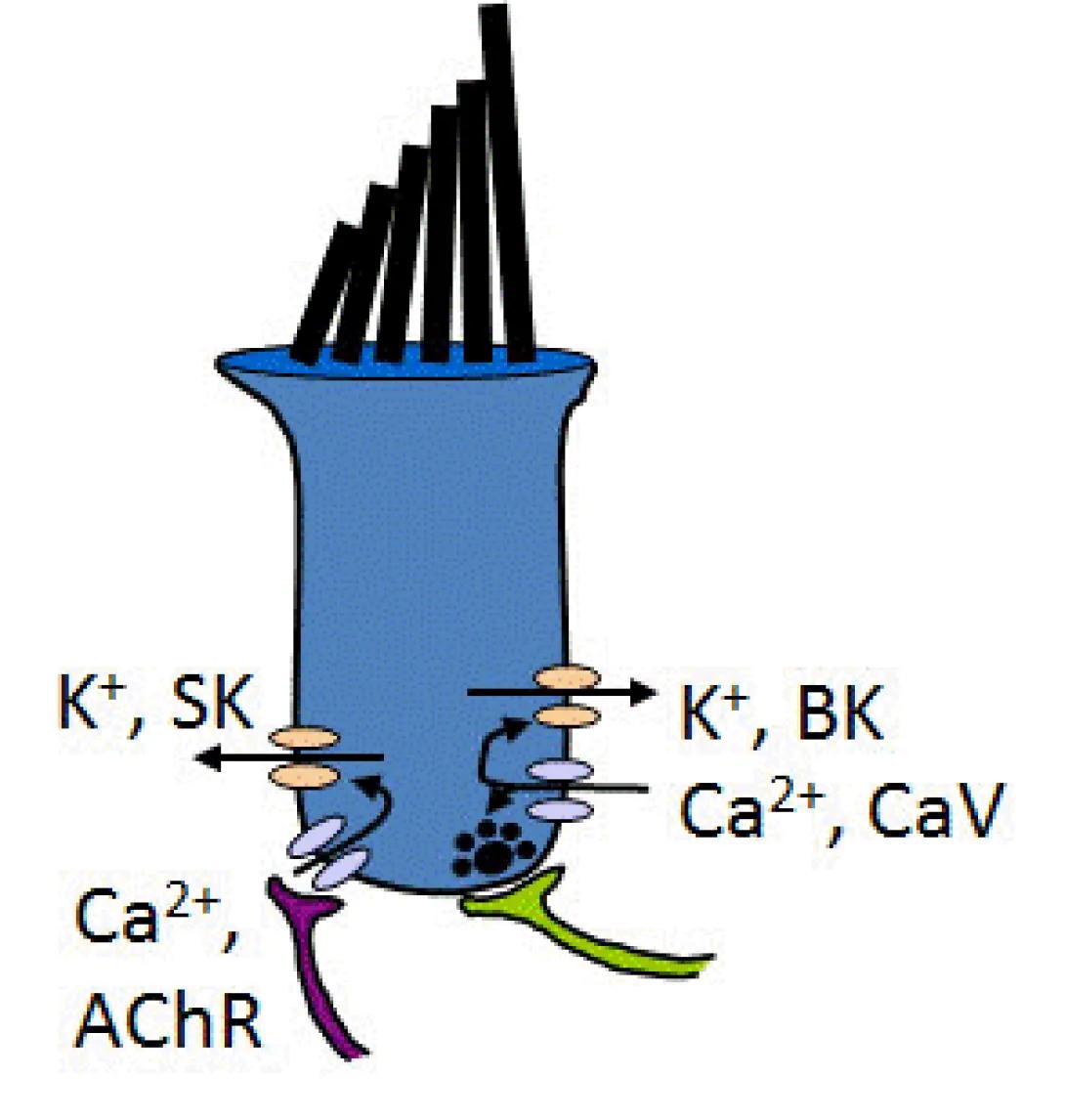
Cochlea hair cells are highly polarized and their membrane processes are exquisitely compartmentalized. The generic hair cell shown here illustrates efferent (SK/AChR) and afferent (BK/CaV) synapses, each of which contain a large number of proteins clustered into discrete domains.
Lipid Rafts and Hearing Loss
In our search for the mechanisms that organize ion channels into discrete compartments in the cell membrane, we uncovered a new ototoxin, that is, a drug that is used therapeutically but carries with it an unfortunate risk to hearing health. This drug is a simple ring of sugars called cyclodextrin (specifically, 2-hydroxypropyl-beta-cyclodextrin, or HPBCD). Cyclodextrins are widely used in biophysics to manipulate cell membranes by mobilizing cholesterol. Originally, we used these compounds to test whether ion channel clustering in hair cells occurred in lipid microdomains. We were surprised to find that cyclodextrin enhanced calcium current while inhibiting potassium current. In Motor City terms, this would be equivalent to stepping on the gas while releasing the brake, a citation that could lead to excitotoxicity in cells. While generally recognized as safe, we found that high doses of HBPCD were capable of rapidly inducing hearing loss in mice by destroying cochlear hair cells. Clinical trials using HPBCD to treat lipid disorders are seeing increased hearing loss risk in human patients. Current projects seek to understand the relationship between cholesterol, excitability and hair cell health in order to gain insight into basic membrane biology and design ways to mitigate the risk to hearing with HPBCD use in the clinic.
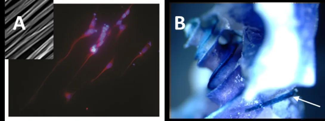
As an example, two hair cells are shown below with antibody stains to the BK-type potassium channel, clustered into hot spots at the base of each cell.
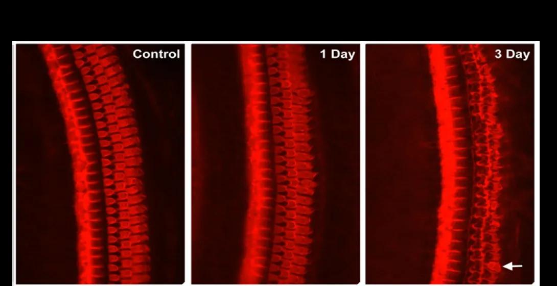
When cyclodextrin (HPBCD) is given subcutaneously to normal mice, we see widespread loss of outer hair cells within 3 days of treatment. Current projects are looking at the mechanisms and mitigation of this devastating effect.
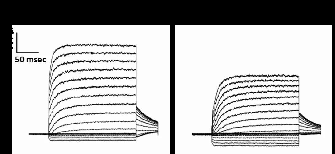
Although the ear appears quite normal at the onset of hearing, the acquisition of sound transduction in the ear represents a massive molecular remodeling of sensory cells, neurons and their synaptic connections. Early studies in our laboratory focused on the advent of BK-type potassium channels in hair cells, as this is a key event in the onset of hearing, transforming immature spiking hair cells to those capable of encoding high frequency sounds. In these studies we identified major splice variants that govern the expression of these channels, but the molecular mechanisms linking channel expression and hearing sensitivity remain unknown. In collaboration with Dr. Sally Camper, we began exploring the role of thyroid hormone in hearing maturation. Congenital hypothyroidism leads to deafness and the critical window for thyroid hormone activity is at the onset of hearing. BK channel activity, among many other processes, is tightly connected to action of thyroid hormone. Below, we highlight one of our active projects in this area:
Thyroid Hormone and the Acquisition of Hearing in Normal and Regenerating Avian Ears
Thyroid hormone is essential to the maturation of hearing but identification of hormone-sensitive processes in hair cells is confounded by pleiotropic effects in the cochlea and elsewhere. Capitalizing on our previous work in the avian ear, which is capable of spontaneous hair cell regeneration, we are embarking on a new line of research examining the role of thyroid hormone in the regenerating ear. Using the goitrogen methimazole, we have developed methods to reduce total T3 levels by 90% in the embryonic chick ear and in posthatch animals. We have also established methods to eliminate 99% of the sensory hair cells in the chick cochlea using repeated doses of streptomycin. Preliminary experiments in embryos support the requirement for thyroid hormone in the maturation of hearing in chicks. Recent data also supports the hypothesis that thyroid hormone is essential to the regenerating ear. These approaches will provide new insights into the factors that control hair cell maturation and may impact our approach to mammalian hair cell regeneration.
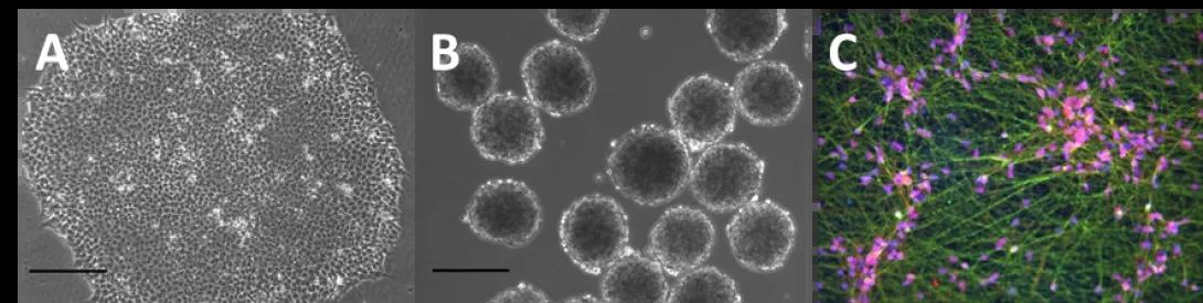
Extraordinary demands of sensitivity and timing are placed on the cochlea, and as a result, small changes in cochlear physiology could greatly impact perception. Sensorineural deficits are often associated with the injury or destruction of sensory hair cells and/or auditory neurons. Therefore, a complete description of their structure and function, throughout development and maturation, is essential to understanding normal behavior, the response to injury and the requirements for regeneration. Two active projects in this area seek to regenerate auditory neurons and sensory hair cells using human pluripotent stem cells. Below we highlight one of our active projects in this area.
Stem Cell Regeneration of Auditory Neurons
Major challenges in stem cell therapy include the need to guide differentiation, guide sub-phenotypes and guide integration with host tissue. With the gracious help of Drs. Sue O’Shea and Rick Altschuler, we ventured into this field using a genetic tool to produce auditory-like neurons. Current projects are translating this work to human embryonic and induced-pluripotent stem cells using a combination of chemical and genetic cues.