Workflows and Standard Operating Procedures, SOPs
Workflows and SOPs
The Workflows and SOPs for each of the 3 Research Cores are available below.
Structural and Compositional SOPs
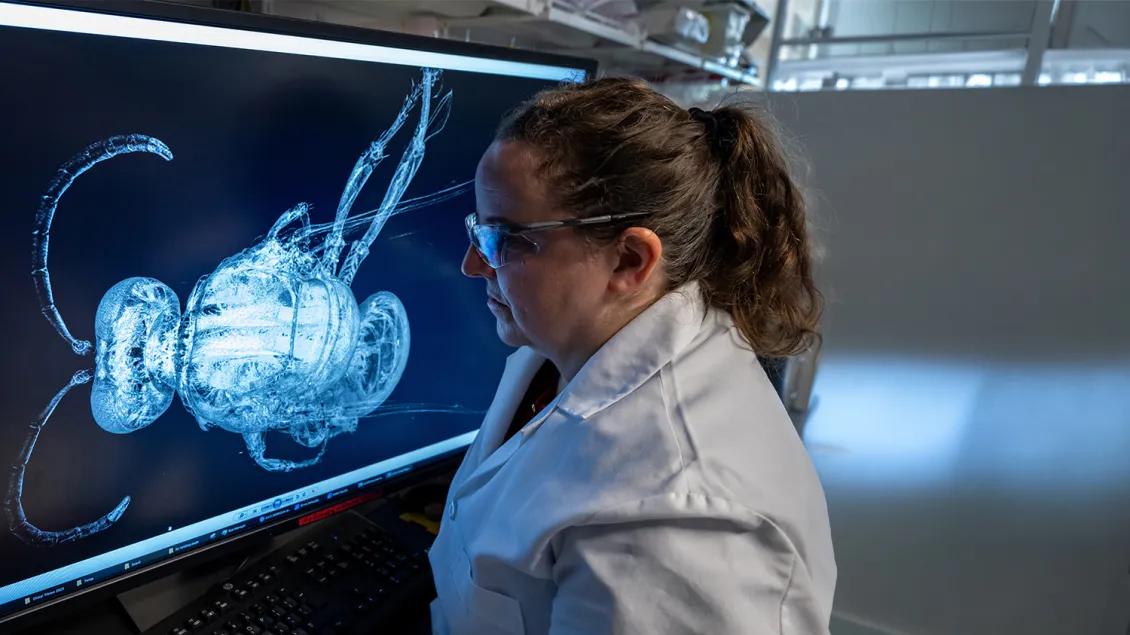
These protocols are designed for a GE phoenix nanotom® s system and are in the process of being updated for the newer GE phoenix nanotom® m system. If you have a different nano-CT system, you will need to adjust the protocol accordingly.
Small animal ex vivo specimen scanning protocols
Protocol for scanning adult mouse femurs
Protocol for scanning adult mouse lumbar vertebrae
Human ex vivo specimen scanning protocols
These protocols are designed for a GE eXplore Locus SP ex vivo preclinical specimen scanner. If you have a different micro-CT system, you will need to adjust the protocol accordingly.
Protocol for setting up and scanning on the eXplore Locus SP scanner
The custom-built Raman microscope features a 785-nm laser and motorized stage, enabling non-destructive, non-invasive, and label-free analysis in point acquisition mode. It can accommodate fresh or dehydrated sections of soft and hard tissues, with polymer-embedding preferred for hard tissues. Hard tissues, whether fresh or polymer-embedded, should be relatively flat and under 2-cm thick to fit on the microscope stage. Additionally, reference compounds, biofluids, and other materials can be analyzed on fused-quartz or alumina-coated slides. Refer to the Raman microscope SOP in the Raman Resource section (below) for safety and operational instructions.
General instrument specifications are outlined below:
- Spectral range: 317 to 1907 cm⁻¹ (fingerprint region for molecular identification)
- Excitation source: 785-nm laser (ideal for biological samples)
- Spectrograph slit width: 25 µm (± 2 cm-1 spectral resolution)
- Detector: Back-illuminated CCD camera (Andor Technologies)
- Objectives: 10x0.50 NA is the default (20x0.75 NA option is available)
- Spatial resolution: 3-5 µm (permits spatial sampling)
The MultiRxn immersion fiberoptic probe from Endress+Hauser features a focus-free design and is constructed from stainless steel with a sapphire window. It requires direct contact with the sample to collect spectra with good signal-to-noise ratios. Samples can be positioned on a manual XY-stage for spectral collection in point acquisition mode. The probe is suitable for non-embedded samples with large surface areas, such as ex vivo human bone tissues and rodent dermal tissue biopsies. It allows real-time in vitro analysis of samples, such as biopsies and biomaterials, in water or buffer. Additionally, we have a custom-made tilted support that allows for the sampling of curved bone surfaces, such as the plates and suture regions of intact whole rodent calvariae. Refer to the Raman fiber-probe SOP in the Raman Resource section (below) for safety and operational instructions.
General instrument specifications are outlined below:
Spectral range: 194 to 1930 cm⁻¹ (fingerprint region)
Excitation source: 785-nm laser (ideal for biological samples)
Spectrograph slit width: 50 µm (± 4 cm-1 spectral resolution)
Detector: Back-illuminated CCD camera (Andor Technologies)
Spatial resolution: ~100 µm (permits bulk or spatial sampling)
This section, offer resources to those using our Raman instruments and provides guidance for individuals interested in exploring their use. We provide laboratory SOPs for our Raman instruments and protocol updates to promote best practices in sample preparation and analysis. Occasionally, we showcase Raman application notes that explore emerging uses in the musculoskeletal and biomaterials research fields.
Onboarding for New Raman Core Users
Laboratory SOP for Raman Microscope
Laboratory SOP for Raman Fiber Probe
Histology Workflows and SOPs
Please discuss your project with a Histologist as far in advanced as possible to ensure you will be able to get your specimens processed correctly and within your project deadlines. All workflows have both self-service and/or Core service components.
Paraffin embedding and sectioning workflow
Cryostat sectioning (non-hazardous)
Cutting and grinding thick plastic sections using Buehler’s Isomet and EcoMet
Freezing and Embedding Tissue for Cryostat Sectioning
Methyl Methacrylate Solutions and PMMA
Methyl Methacrylate Processing and Embedding
Sectioning Paraffin Blocks, Automated
Sectioning Paraffin Blocks, Manually
Toluidine Blue for Plastic Sections of Soft Tissue (Thick)
Toluidine Blue-Basic Fuchsin for Plastic Sections (Thick)
The "Bone Histomorphometry Booklet" contains all the protocols relative to plastic thin sectioning and histomorphometric analyses used by the P30 Histology Assessment Core. In this booklet, you will find information on preparing and submitting specimens to the Core, embedding the specimens and sectioning, staining the sections, acquiring microscope images of the sections, and histomorphometric analysis of the images.
Karl Jepsen, PhD ([email protected])
Carol Whitinger ([email protected])
Functional SOPs
The Animal Phenotyping and Functionality Core recommends designing and conducting whole-animal and muscle phenotyping studies based on the Standard Operating Procedures published by the TREAT NMD Group for studying mouse models of neuromuscular disease. These protocols are validated and well accepted in the field for phenotyping and standardizing preclinical testing. Modest deviations from the protocols may be required to accommodate differences in equipment within the core. Please consult the core directors regarding any training/consultation on how the core's equipment is used, and any required deviations from these SOPs.
Use of treadmill and wheel exercise for impact on mdx mice phenotype
Use of grip strength meter to assess the limb strength of mdx mice
The use of hanging wire tests to monitor muscle strength and condition over time
Behavioural and Locomotor Measurements Using Open Field Animal Activity Monitoring System
Use of treadmill and wheel exercise to assess dystrophic state
Whole femur bending tests
- Mouse 4 point bending protocol. This protocol was written for use with a custom testing frame used on an MTS mini-bionix system.
- Four point bending for human long bones. This protocol was designed for use with custom fixtures on an Instron testing frame.