
Radiation Oncology 3D Print Lab
The Radiation Oncology 3D Print Lab accelerates the development and fabrication of patient-optimized treatment aids and research tools.
Our dedicated 3D part design and manufacturing lab assists Radiation Oncology to explore cutting edge research and further improve our patient care. High quality 3D printing using a wide variety of materials allows for rapid collaborative progress at the point-of-care.
For More Information
Current Projects
Each component is manufactured with resins optimized for its specific application. With rapid 3D prototyping, the path from inspiration to application has never been shorter.
Medical Procedure Aids
- Needle Fixtures and Guides (Brachytherapy)
- Radiation Source Templates (Brachytherapy)
- Radiation Blockers (Boluses, Lung Blocks)
- Patient Aids (TSET Stand, Angled Patient Chair)
Quality Assessment Tools
- Geometric and Dosimetric Phantoms (Winston Lutz Phantoms, RGSc System Phantoms)
Specialty Laboratory Apparatus
- Small Animal Research Aids
- Gel Electrophoresis Combs and Molds
Examples of Clinical Projects
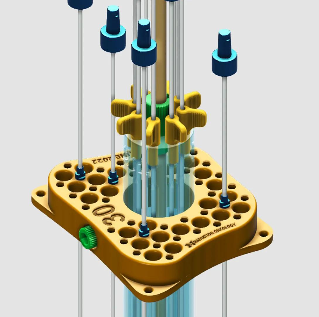
GYN Brachytherapy Applicator:
Patient-specific interstitial cylinder and plate for gynecological brachytherapy procedure (in-situ for up to 60 hours). Printed with biomedical resin approved for steam sterilization and extended contact with skin and mucosal membranes.
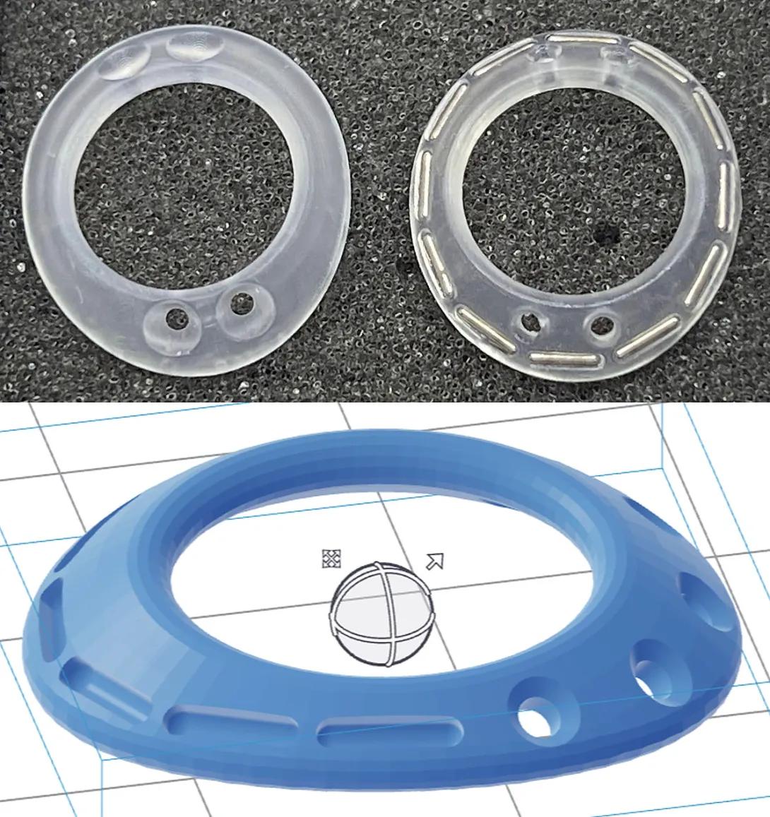
Opthalmic Radiotherapy Plaque:
Patient-specific eye plaque conformer for brachytherapy procedure. Printed with biomedical resin.
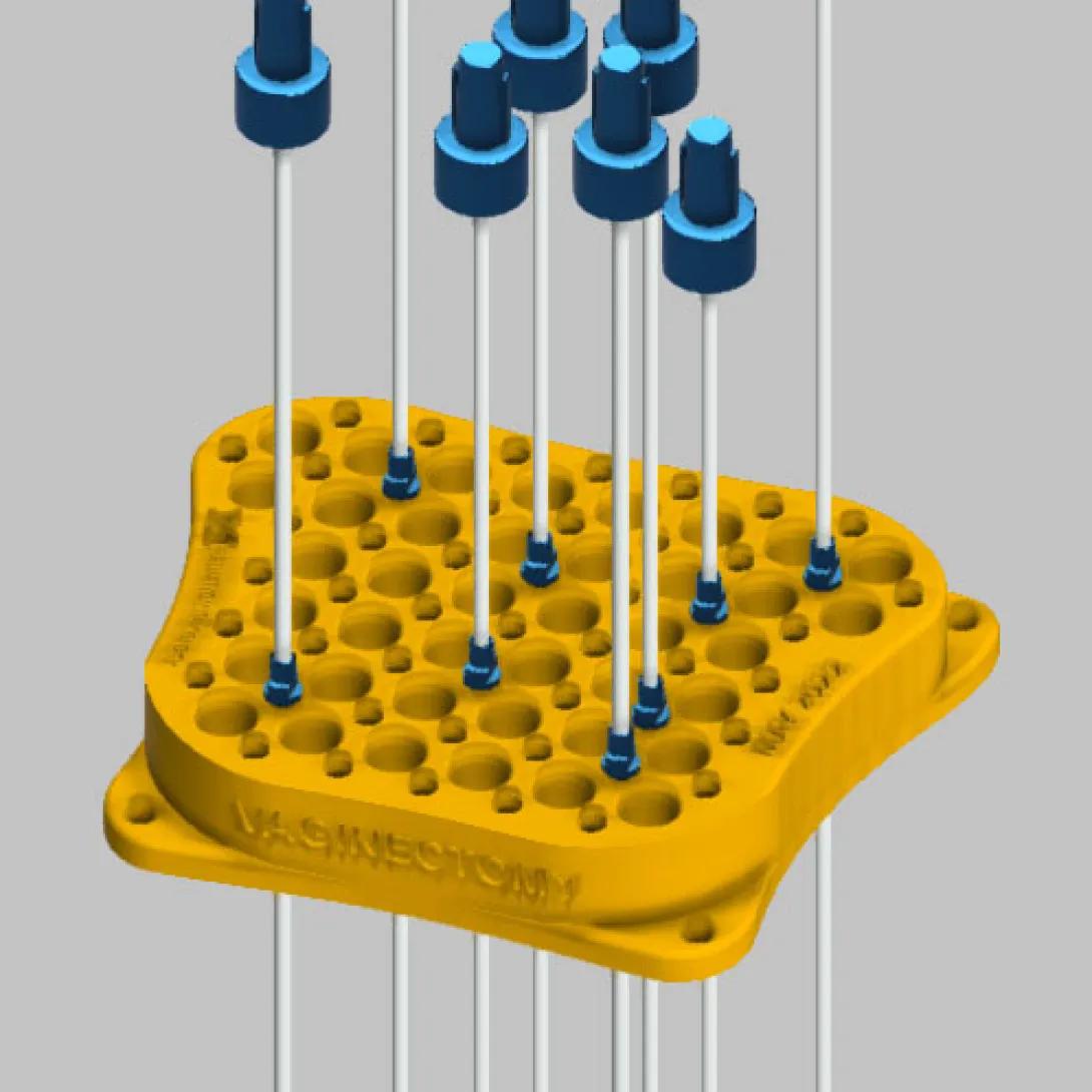
Brachytherapy Needle Guide:
Custom-sized plate printed with approved biomedical resin for brachytherapy treatment.
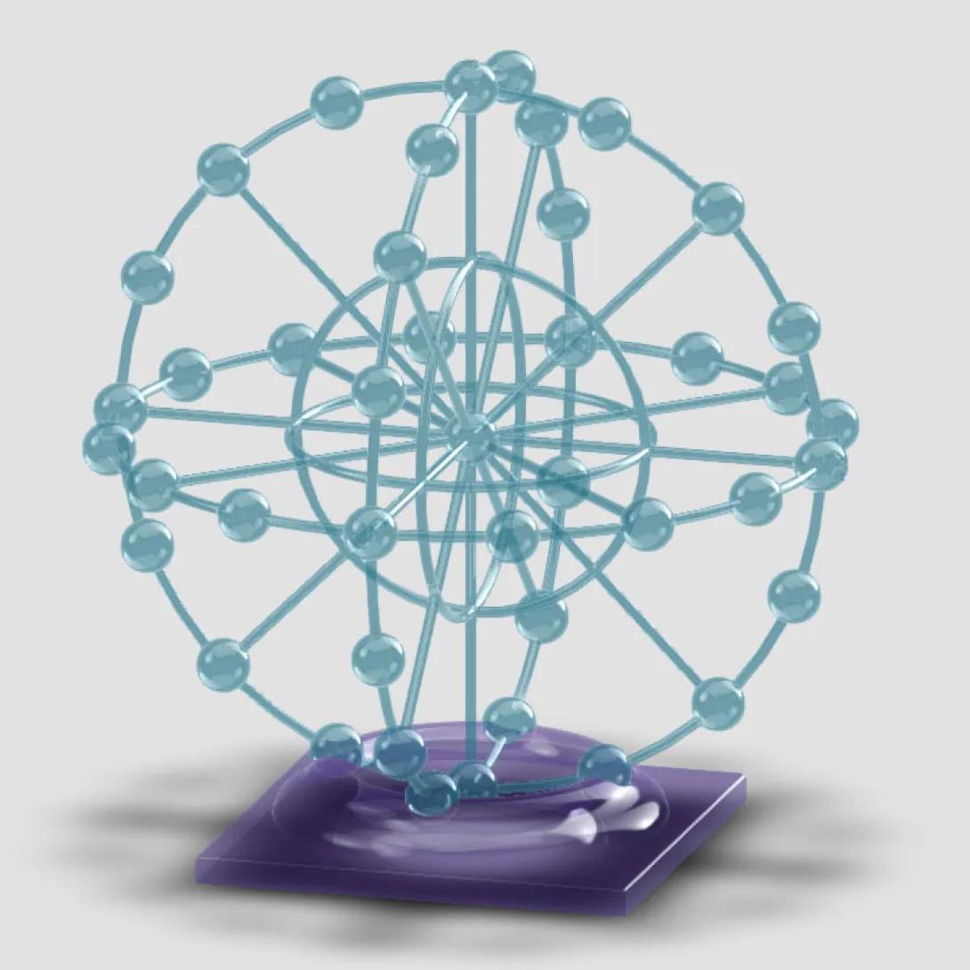
Phantoms for precision treatment calibration:
3D spherical array
Radiation beam blockers:
Tissue-sparing lung block prototype customized for each patient from DICOM data. Blocks are filled with reusable metal bearings.
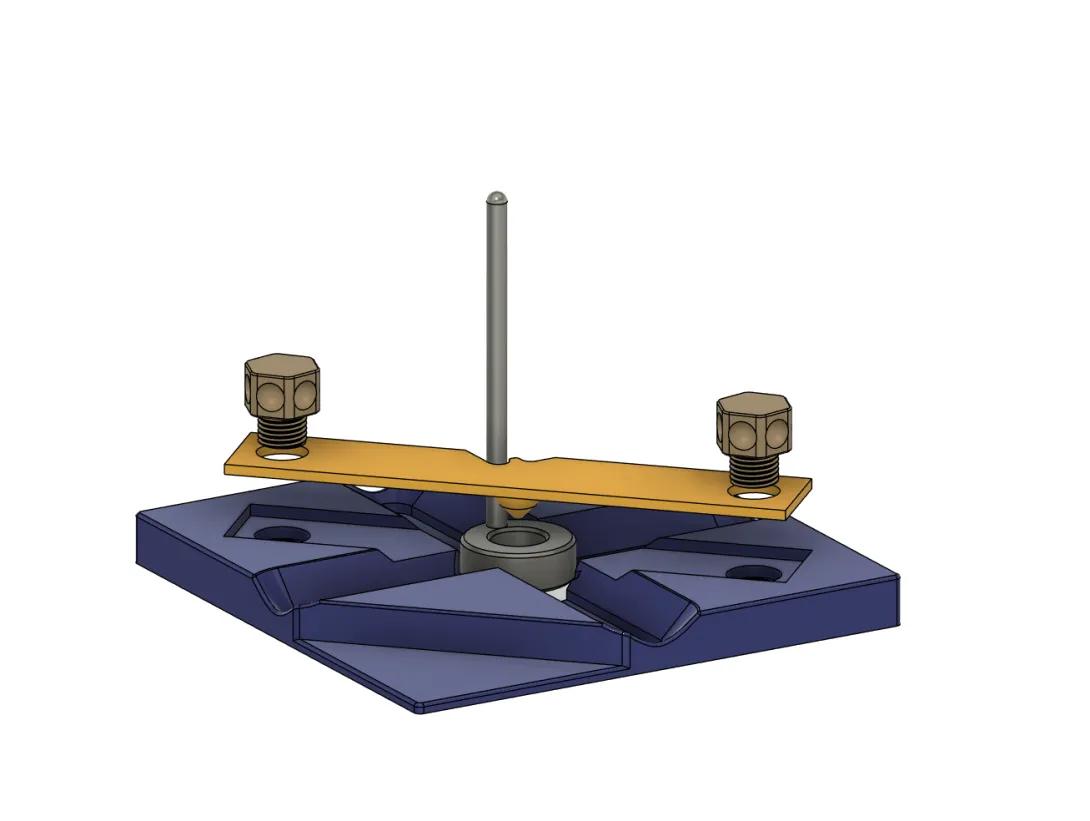
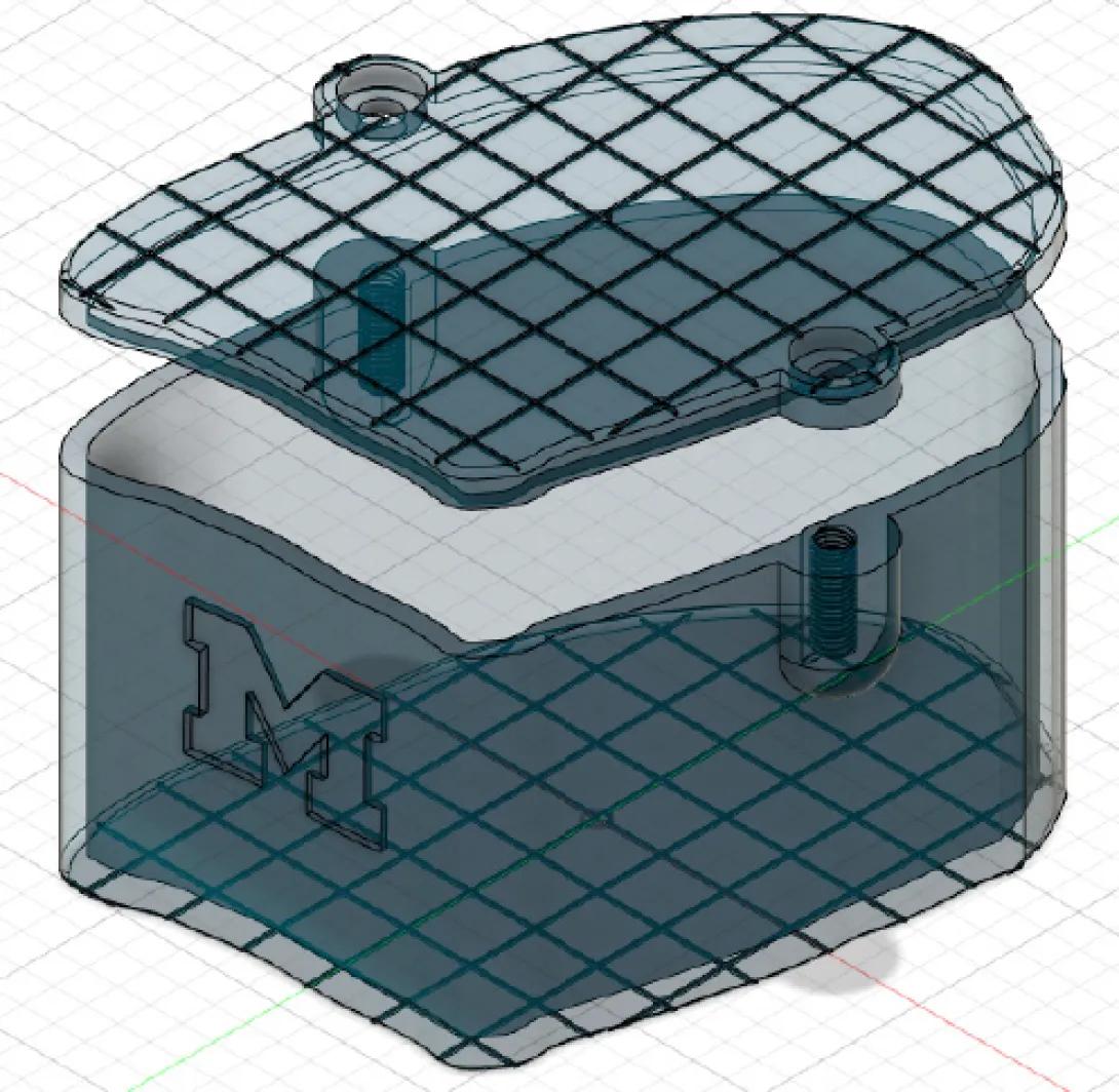
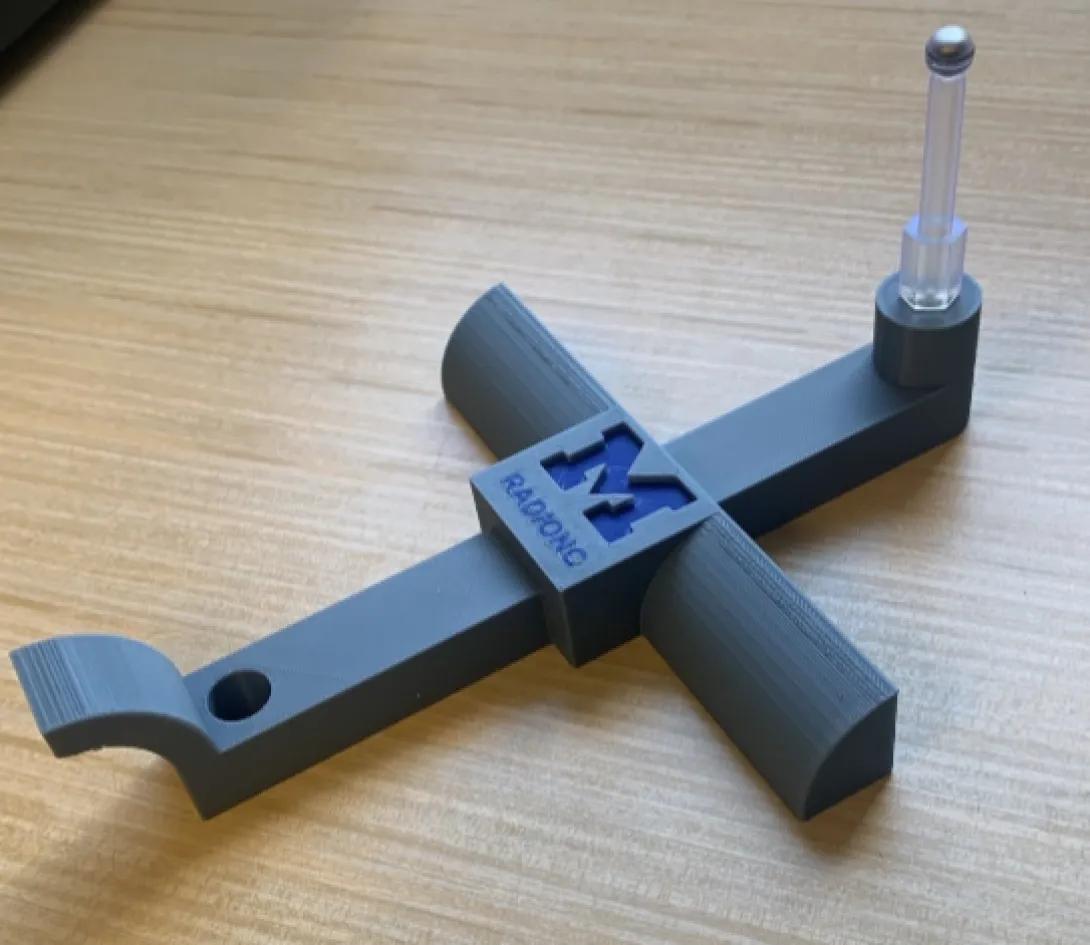
Ring Applicator Stand:
Repeatable fixture for quality assessment of Brachytherapy devices.
Winston Lutz Phantoms:
Quality assessment tools made to fit specific patient treatment machines.
Examples of Laboratory Projects
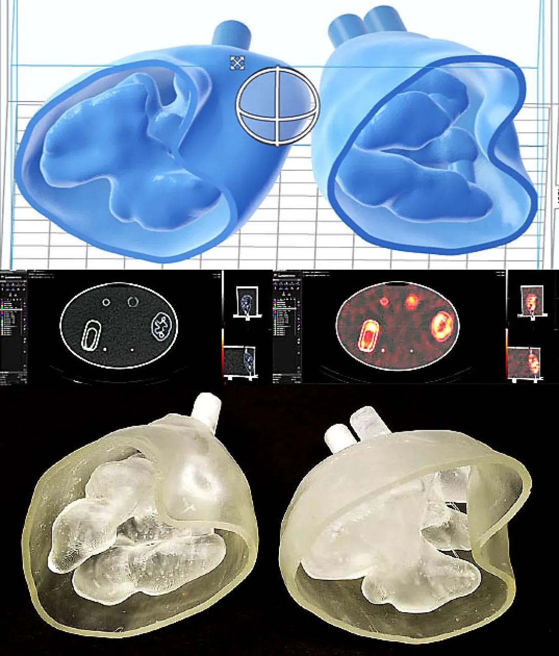
Simulated organs for translational research:
Images of the 3D printed kidney phantom (printed in two halves and glued together) with separately fillable cortex and medulla, and modeled from a DICOM image from the patient. The medulla was filled at 1/3 rd the cortex Lu-177 activity concentration to mimic what is believed to occur in patients undergoing Lu-177 DOTTATE therapy.
Phantoms for research:
Transducer model to take ultrasound images concurrent with radiation therapy.

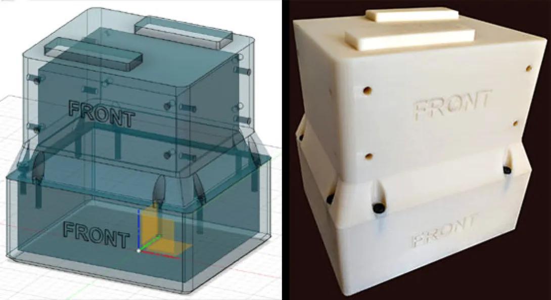
Specialty laboratory equipment:
Corner supports to suspend a polycarbonate shield beneath the Orthovoltage unit.
3D Service Team

Steven Kronenberg
3D Service Manager

Choonik Lee, PhD
Clinical Professor of Radiation Oncology
Medical School
Charles Matrosic, PhD
Medical School

Alexander Moncion, PhD
Program Director, Radiation Oncology, Medical School
Kwok Lam, PhD

Marc L Kessler
Professor Emeritus of Radiation Oncology
Clinical Professor Emeritus of Radiation Oncology
Medical School