Research | Fort Lab
Research
Overall, the Fort lab focuses on increasing our understanding of retinal physiology, and how it is affected by neurodegenerative and neuroinflammatory diseases such as Diabetic Retinal Disease, the main ocular complication associated with diabetes. Neurons and glia within the retina interact continuously to support visual processing and maintain a healthy tissue. Most recently, two major non-neuronal cells are of particular interest to us: Müller glia, they span the entire retinal thickness, providing metabolic and trophic support, regulating extracellular ions and neurotransmitters recycling, regulating tissue homeostasis including through modulation of tissue inflammation, and coupling to neurons through signaling pathways that stabilize synaptic activity. Microglia survey the retinal environment and communicate with neurons to modulate synaptic remodeling and respond to injury or inflammation, helping preserve overall retinal homeostasis.
For many years, Dr. Fort’s laboratory has been using non-targeted discovery approaches to expand our understanding of disease pathophysiology in an unbiased manner. This approach started with the use of multi-omics analysis of retinal tissues from animal models of diabetes and led to the novel discovery of the induction of proteins of the crystallin family (Fort et al. 2009). Among these proteins, the Fort lab has particularly focused on the role and regulation of one of its members: alphaA-crystallin. AlphaA-crystallin belong to the small heat shock proteins (sHSPs or HSPBs) and represent a critical intrinsic mechanism of cell survival in response to a variety of stress conditions and that their function is dramatically affected under chronic conditions such as diabetes (Ruebsam et al,. 2018). Most recently work from multiple trainees and staff members in the Fort lab demonstrated that this pathway plays a central role in regulating neuroinflammation, especially through its expression and secretion by the main glial cells of the retina (Nath, et al. 2021). We further characterized its mechanisms of regulation (Nath et al. 2022a and Sluzala et al. 2024) but also its therapeutic potential in multiple cell and animal models of retinal neurodegenerative conditions (Wang et al, 2021, Nath et al. 2022b, Besirli et al. 2024). The current research of our laboratory focuses on:
Role of HSPB4/CRYAA in the regulation of the multiple function of the Müller glial cells in normal and pathological conditions.
Using a combination of cell and animal models, the goal is to define how HSPB4/CRYAA is involved in the regulation of the multimodal supportive role of Müller glial cells under normal, but also metabolic stress, acute and chronic. Our current research explores the endogenous role of HSPB4/CRYAA as well as the impact of its supplementation on the stress-induced gliotic response of Müller glial cells.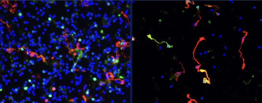
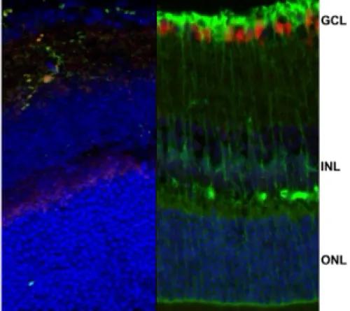
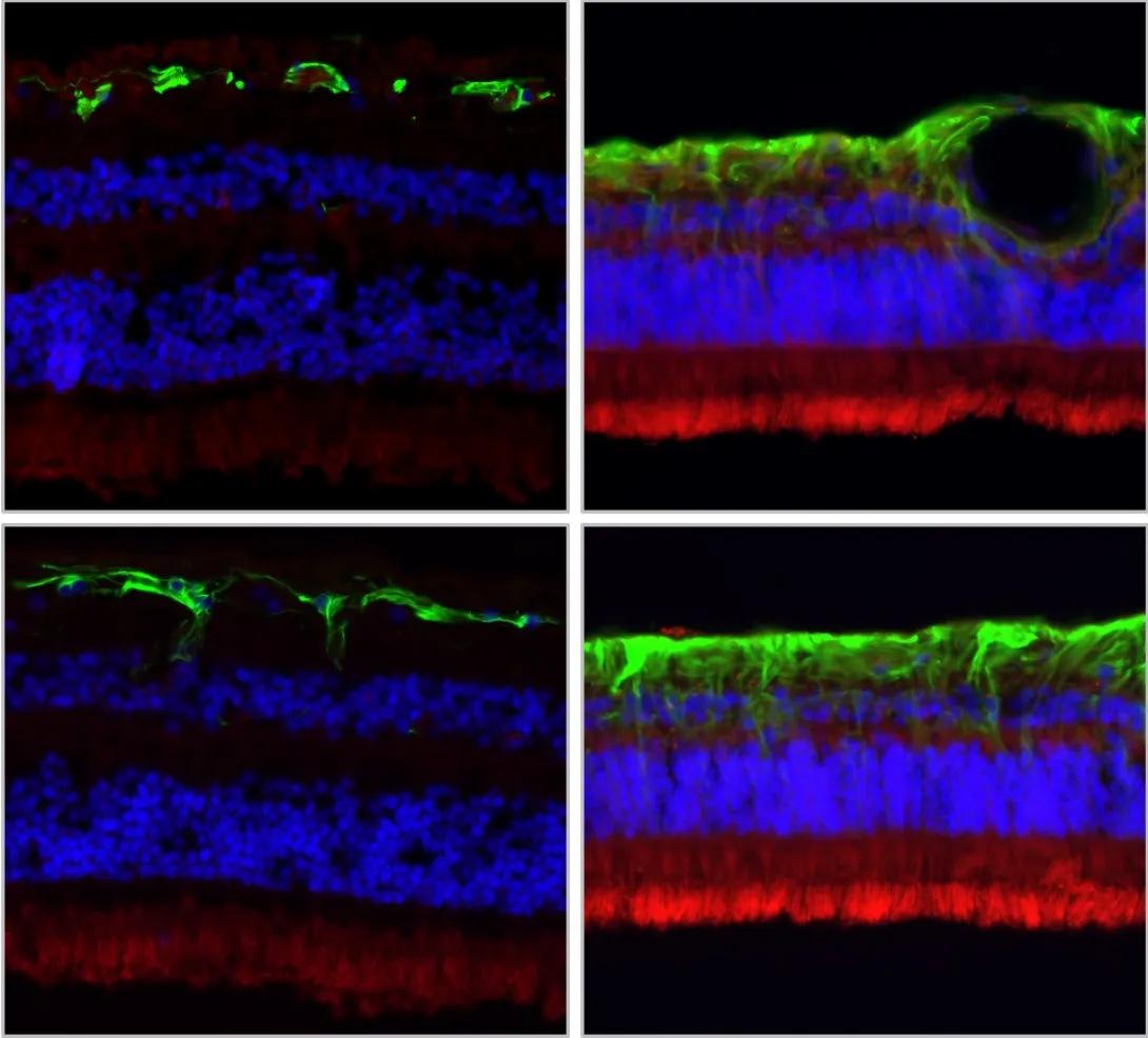
Representative images of dissociated retinal cells before (left) or after flow cytometry based isolation of GFP (green) positive Müller glial cells confirmed by IF staining for a specific cell marker (red). (blue is a marker of cell nuclei) 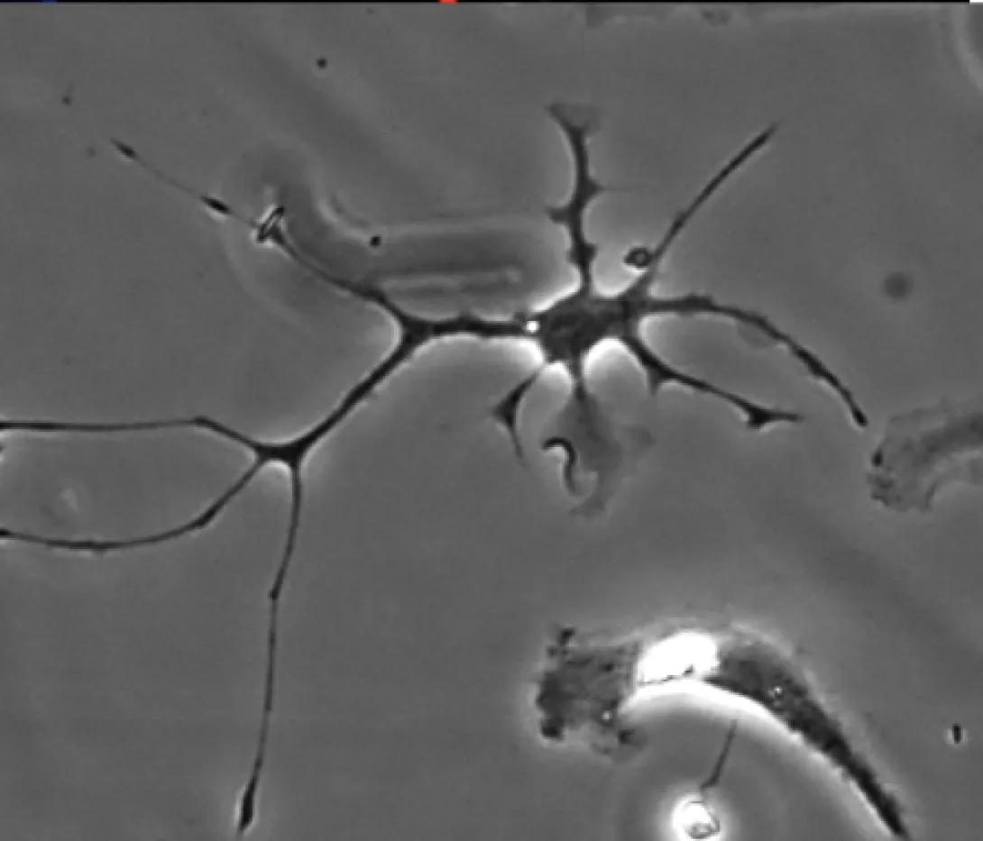
Role of HSPB4/CRYAA in the regulation of the stress-induced activation of microglial cells. Müller glial cells and microglia both play critical roles in sensing and modulating the retinal homeostasis to promote proper retinal function. Both cell types are key partners that depend on each other and led us to ask if HSPB4/CRYAA produced and secreted by Müller glial cells could play a key role in regulating stress-induced microglial activation.
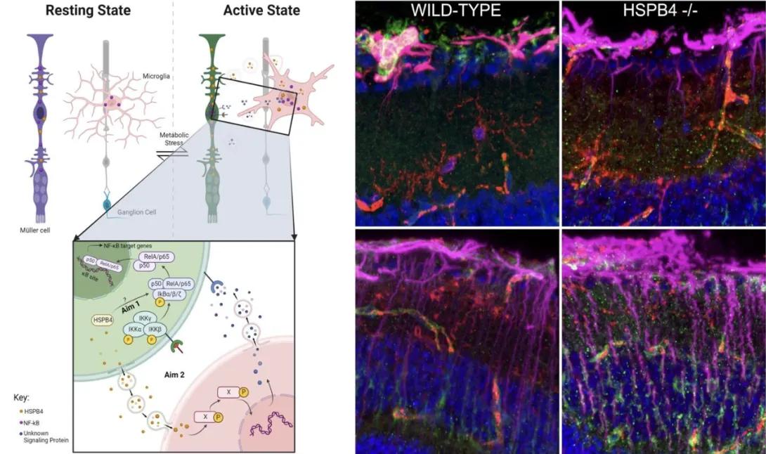
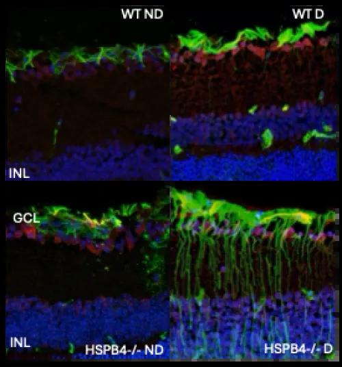
Schematic of the current working model relative to the interplay between Müller glial and microglial cells and its modulation by HSPB4 under normal and disease conditions (left). Representative images of retinal cross-sections from mice expressing (WT) or not HSPB4 (HSPB4-/-) with (right) or without diabetes (left) and stained for a marker of gliosis (purple), a marker of microglia (red) and a marker of inflammation (green).
The second main goal of Dr Fort’s laboratory is to define the molecular mechanisms of the impact of diabetes on the retina in humans to better our understanding of human conditions and help increase the translatability of research findings obtained from cell and animal models. One of the unique characteristics of the retina is that it is virtually the only tissue in the body that cannot be biopsied. Human studies require close collaboration with organ procurement organizations and eye-banks to establish a much-needed biorepository of ocular tissues from human donors. This was made possible due to the selfless and generous contributions of donors and their families, and the hard work of the above-mentioned key organization and their dedicated staff. This work was initiated in conjunction with the use of state-of-the-art discovery approaches, which first helped demonstrate the impact of diabetes on retinal lipid synthesis and the alteration of fatty acid beta-oxidation, a mechanism critical for cell survival and proper retinal physiology and likely central to the progression of diabetic retinal disease (Fort et al, 2021).
In addition to enabling state-of-the-art discovery approaches that are progressively uncovering novel molecular mechanisms of disease pathophysiology, these ocular tissues have also been central to validating pathways identified through other means, helping confirm their relevance in the human context (Ruebsam et al, 2018, Liu et al, 2022, Liu et al, 2023, Lin et al, 2025).
This work has inspired the establishment at the Kellogg Eye Center of the Mary Tyler Moore Vision Initiative Ocular Biorepository and Resource Center (MTM Vison BRC) for which Dr Fort is the director. In addition to having already established the largest and most deeply characterized collection of ocular tissues from human donors with diabetes, the MTM Vision BRC is generating large multi-omics datasets characterizing these tissues and linking them to the first web-based platform of its kind to be used by the scientific community involved in the consortium.