Razumilava Lab Research
Learn more about our work and impact.
Our group promotes innovation, rigor, and inclusivity of science. Our ultimate goal is to improve outcomes for patients with biliary disorders through scientific discoveries.
The extrahepatic bile duct (EHBD) is an understudied structure whose disease outcomes, like cholangiocarcinoma, are associated with poor prognosis. With limited treatment options, the goal of the Razumilava lab is to understand the natural mechanisms of proliferation for recovery and how these mechanisms become pathogenic responses in cancer development.
Current Projects
EGFR activation in cholangiocytes promotes extrahepatic bile duct regeneration after injury.
Calder et al., Hepatology Communications. 2025. PMID: 40568138.
This study investigated the role of EGF family signaling in the cholangiocyte proliferative response to biliary obstruction. Several EGF family ligands are upregulated post-injury activating EGFR and inducing proliferation.
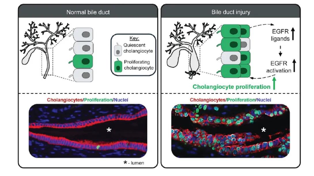
This image compares the state of cholangiocytes in normal and injured bile ducts. In normal conditions (left), most cholangiocytes are quiescent (gray), with very few proliferating cells (green), as shown in both the schematic and the histology image (red = cholangiocytes, green = proliferation, blue = nuclei). Upon injury (right), EGFR ligand expression and activation increase, leading to a significant rise in cholangiocyte proliferation (more green cells), which is evident in both the schematic and the corresponding histological image. This highlights that bile duct injury stimulates cholangiocyte proliferation through EGFR signaling to promote repair.
WNT signaling contributes to the extrahepatic bile duct proliferative response to obstruction in mice.
Calder et al., JCI Insights. 2024. PMID: 39636699.
This study uses mouse and human organoids and mouse explants to investigate WNT activation and inhibition. WNT inhibition decreased cholangiocyte proliferation to acute obstruction. Cholangiocytes were shown to be both a source and target of WNT signaling.
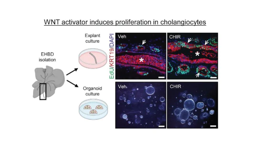
This image demonstrates that WNT activation increases proliferation in cholangiocytes. The schematic on the left shows EHBD (extrahepatic bile duct) isolation followed by either explant culture (top) or organoid culture (bottom). On the right, histological images compare vehicle-treated (Veh) and WNT activator-treated (CHIR) samples. In explant cultures, EdU (green) marks proliferating cells, KRT19 (red) labels cholangiocytes, and DAPI (blue) stains nuclei; more green cells (arrows) are seen in CHIR-treated than vehicle, indicating increased proliferation upon WNT activation. The asterisks mark the bile duct lumen. In organoid cultures, more and larger organoids are visible with CHIR treatment compared to vehicle, further showing enhanced proliferation and growth with WNT activation.
C-X-C motif chemokine ligand 1 induced by Hedgehog signaling promotes mouse extrahepatic bile duct repair after acute injury.
Mohamad Zaki et al., Hepatology, 2022, PMID: 35388502.
This study revealed that Hedgehog signaling regulates CXCL1 which in turn directs the inflammatory response and biliary proliferation after acute extrahepatic bile duct injury. This is shown by decreased CXCL1 expression in genetically inhibited Hedgehog mice (Gli1-/-) or chemically inhibited mice (LDE) corresponding with decreased neutrophil recruitment (Lys6G+). Then connecting the two ideas, mice were treated with a chemical inhibitor (SB) of neutrophil recruitment which corresponded with decreased epithelial cell proliferation.
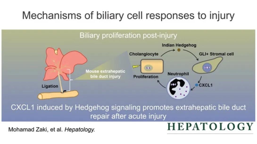
This image illustrates the mechanism by which biliary cells respond to injury, focusing on the process of biliary proliferation after damage. It shows that in a mouse model, extrahepatic bile duct injury is induced by ligation. Following injury, cholangiocytes (bile duct cells) produce Indian Hedgehog, which signals to GLI-positive stromal cells. These stromal cells then release the chemokine CXCL1, which attracts neutrophils to the site of injury. Neutrophil recruitment and the resulting signaling promote cholangiocyte proliferation. The key message is summarized below: "CXCL1 induced by Hedgehog signaling promotes extrahepatic bile duct repair after acute injury." This mechanism highlights the importance of immune and signaling pathways in tissue repair following bile duct injury.
Hepatobiliary Organoids and Their Applications for Studies of Liver Health and Disease: Are We There Yet?
Shiota et al., Hepatology, 2021, PMID: 33638203.
This review summarizes available adult cell-derived intrahepatic biliary, extrahepatic biliary, and gall bladder organoids and their applications. This technology is in its infancy, so some challenges it faces is co-culture development to better model in vivo tissue structure, the imperfections of extracellular matrices, and the lack of standardized protocols. However, the organoids enable studying of models at homeostasis and in disease.
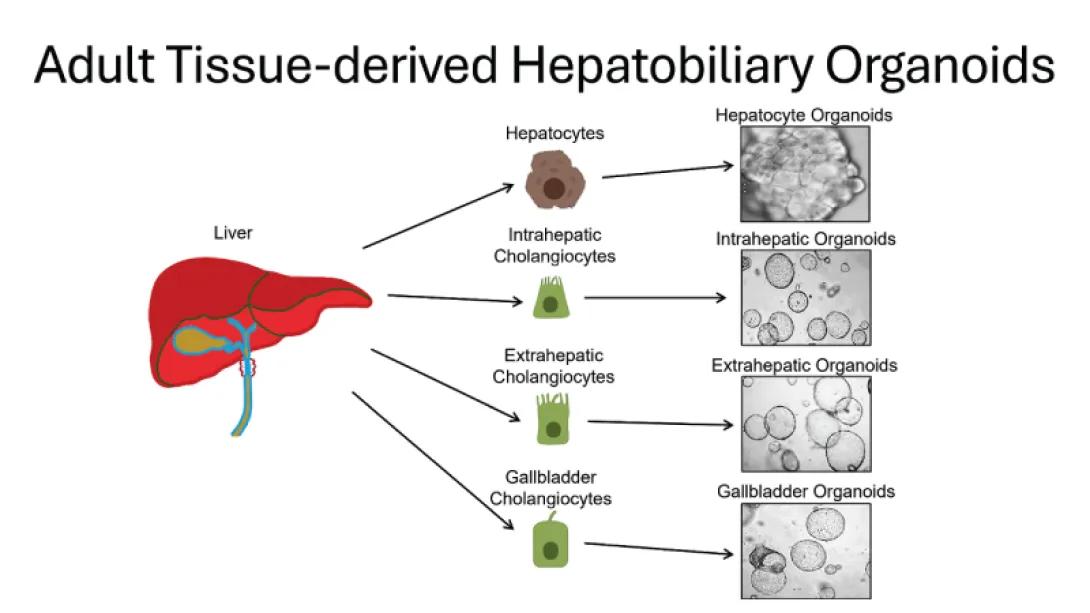
This image shows how different cell types from adult liver and biliary tissue can be isolated to generate organoids in culture. On the left, a diagram of the liver highlights the tissue sources: hepatocytes, intrahepatic cholangiocytes, extrahepatic cholangiocytes, and gallbladder cholangiocytes. Arrows indicate that each cell type can be cultured to form its corresponding 3D organoid: hepatocyte organoids, intrahepatic organoids, extrahepatic organoids, and gallbladder organoids, all shown in microscope images on the right. This illustrates the diversity and potential of adult tissue-derived hepatobiliary organoids for research and regenerative medicine.
Generation of Organoids from Mouse Extrahepatic Bile Ducts.
Shiota et al., J. Vis. Exp., 2019, PMID: 31081815.
EHBD organoid development from neonatal or adult mice consist of mainly cystic morphology through all passages with rare irregular organoids. These cells show markers of E-cadherin, PDX1, CK19, and Sox9 which are markers for epithelial cells, biliary progenitor cells, and biliary differentiation respectively. This protocol allows for continuous study of mechanisms of EHBD cholangiocyte homeostasis as well as future applications to study disease states, genetic, pharmacologic, or infectious disease treatment.
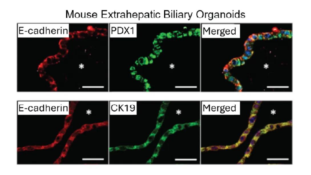
This image shows immunofluorescence staining of mouse extrahepatic biliary organoids. The top row displays organoids stained for E-cadherin (red), PDX1 (green), and their merged image, illustrating that many cells co-express these markers. The bottom row shows organoids stained for E-cadherin (red), CK19 (green), and their merged image, again showing co-expression. The asterisks (*) indicate the lumen of the organoid structures. These results demonstrate that the organoids consist of cells expressing key biliary and epithelial markers (E-cadherin, PDX1, CK19), confirming their extrahepatic biliary identity.
Hedgehog Signaling Modulates Interleukin-33-Dependent Extrahepatic Bile Duct Cell Proliferation in Mice.
Razumilava et al., Hepatology Communications, 2018, PMID: 30766964.
This study shows that inflammatory cytokine (IL)-33 and Hedgehog (HH) signaling are synergizing components to Extrahepatic Biliary proliferation. Treatment with (IL)-33 induced proliferation in all mouse models; WT, pCMV-Shh model which overexpresses HH signaling, and Gli1 LacZ/LacZ model which lacks HH signaling. The results suggest IL-33 primarily drives extrahepatic bile duct (EHBD) cell proliferation because when HH signaling is present, the proliferation rate of the cells is higher than when it is absent. Thus suggesting a synergetic increase in proliferation between IL-33 and HH signaling.
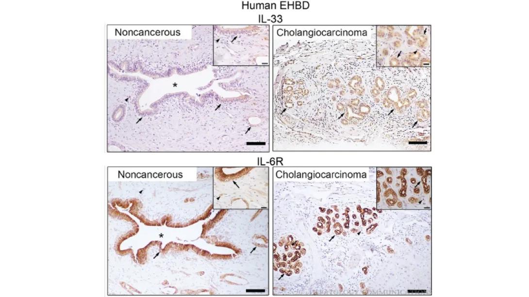
This image shows immunohistochemistry staining of human extrahepatic bile duct (EHBD) tissues for IL-33 (top row) and IL-6R (bottom row), comparing noncancerous tissue (left panels) to cholangiocarcinoma (right panels). In noncancerous tissue, the bile duct epithelium lining the lumen (asterisk) shows weak or patchy IL-33 (top left, arrows) and IL-6R (bottom left, arrows) staining. In contrast, cholangiocarcinoma samples display strong and widespread IL-33 (top right) and IL-6R (bottom right) staining in tumor cells (arrows), as well as insets highlighting cellular detail. These results suggest upregulation of IL-33 and IL-6R in cholangiocarcinoma compared to noncancerous EHBD tissue.