
Nano Computed Tomography Advanced Imaging Core
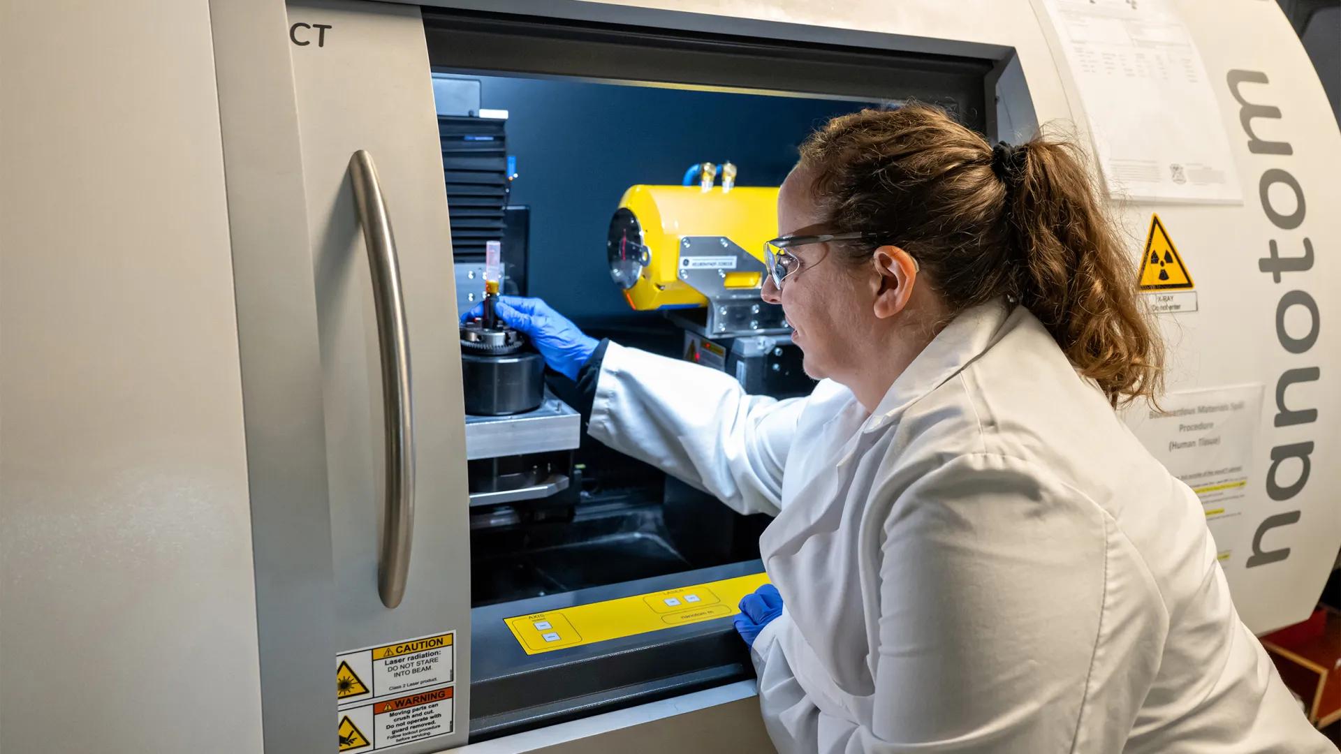
Welcome To The Nano Computed Tomography Advanced Imaging Core
The nano-CT facility core provides services for acquiring three-dimensional imaging and quantitative analysis of biological and engineered structures/materials. The core is capable of imaging mineralized tissues (e.g., bone, teeth) and soft tissues (e.g., tendons, ligaments, cartilage, blood vessels, muscle) with the aid of radiographic contrast agents. This core is managed and operated by members of the Orthopaedic Research Laboratories. Explore the website to see what we can do and to learn about projects under development. Please also check out the MiMHC Education page for training offered on using Dragonfly3D to analyze your nanoCT images.
For general inquiries and specific requests, please contact Andrea Clark at [email protected].
If utilizing the ORL Nano-CT core, please include the appropriate Research Resource Identifier (RRID) when citing the resource:
University of Michigan ORL Nano-CT Core RRID:SCR_026772
High Resolution Bone Imaging
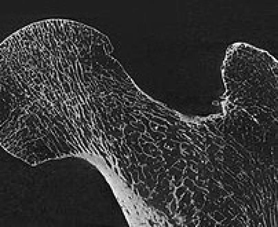
High resolution imaging of ex vivo and in vivo biological materials.
Soft Tissue/Contrast Enhanced Imaging
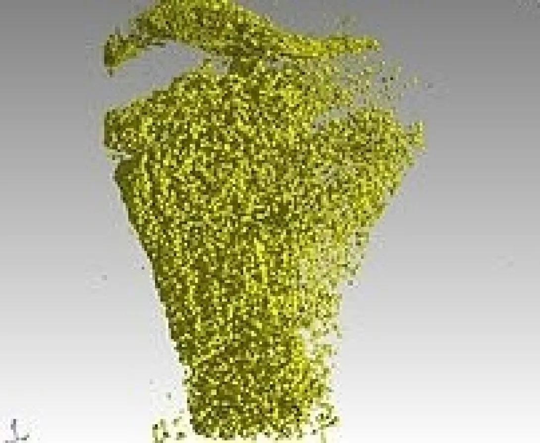
Imaging of soft tissues can be achieved with the use of contrast agents. Structures and organs such as muscles, vessels, heart, kidney, cartilage and others can be imaged.
Beyond Bone...

High resolution imaging of other materials is possible with the nano-CT system, such as metals, ceramics, scaffolds, insects, plants, etc.
Contact Us
2800 Plymouth Road, Ann Arbor, MI 48109, United States
Contact Andrea for assistance with scans, reconstructions, image analysis, and protocol development.
2800 Plymouth Road, Ann Arbor, MI 48109, United States
Contact Dr. Jepsen for assistance with developing new research projects involving nano- or micro-CT imaging.
