
Services | Nano CT Advanced Imaging Core

Services
As part of the Structural and Compositional Assessment Core in the Michigan Integrative Musculoskeletal Health Core Center (MiMHC), the Orthopaedic Research Laboratories nano-Computed Tomography facilities provide full 3-D reconstructed images of hard and soft biological tissues, as well as non-biological materials in conjunction with the MicroCT Core in the Dental School. The Orthopaedic Research Laboratories have the ex vivo GE phoenix nanotom m nanoCT scanner.
ex vivo Scanning
nanotom m
The Phoenix | NanoTom M is an ex vivo nanofocus computed tomography system capable of achieving resolutions down to 300nm (0.3 µm), depending on specimen size. It has a new open 180 kV / 15 W high-power nanofocus X-ray tube with down to 200 nm detail detectability, optimized for long-term stability. Unique temperature-stabilized digital GE DXR detector (3,072 x 2,400 pixels) with a high dynamic range > 10,000:1 and up to 4 times faster data acquisition at the same image-quality level. The maximum specimen size is 250 mm in height and 250 mm in diameter. Two targets are available, along with a wide selection of filters.
Standard morphological analyses of cortical and trabecular bone are available. These include measurements such as total volume, bone volume, trabecular thickness, trabecular separation, number of trabeculae, structural model index, cortical thickness, cortical inner and outer perimeter, marrow area, and many others. Standards are used to calibrate the CT reconstruction attenuation values to density (Hounsfield Units), thus allowing for bone mineral density measurements as well.
Through the MiMHC P30 core, we are offering training sessions on using Dragonfly3D to analyze your nanoCT images. Please visit the MiMHC Education page to sign up for a training session.
The ex vivo system is available at the same cost of $58.00/hour for acquisition and $58.00/hour for analysis. For MiMHC P30 members, 20% of the cost is covered by the core center for the ex vivo systems. The total cost/sample for scanning and acquisition will depend on the protocol and expected outcome of the image volume. We have many protocols at the ready to achieve outstanding images and have adjusted the scan times while maximizing image quality to keep our costs reasonable. Please contact us for a quote.
ex vivo Scanning Prices
| Service | Procedure | Core Charges (non-members) | P30 Member Cost |
| nanoCT | Acquisition by Core | $61.00/hour | $48.80/hour |
| nanoCT | Analysis by Core | $61.00/hour | $48.80/hour |
| nanoCT | Expediated Acquisition by Core | $122.00/hour | $122.00/hour |
| nanoCT | Expediated Analysis by Core | $122.00/hour | $122.00/hour |
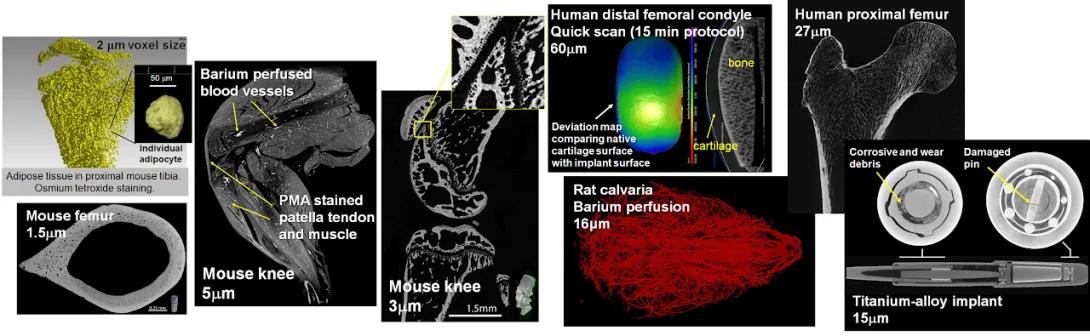
Composite of micro-CT and imaging scans showing bone, vascular, and implant structures in mice, rats, and humans at various resolutions.
• Top left: 3D rendering of adipose tissue in the proximal mouse tibia with osmium tetroxide staining. Inset shows an individual adipocyte at 2 μm voxel size.
• Bottom left: Transverse micro-CT scan of a mouse femur with 1.5 μm resolution. Scale bar 0.25 mm.
• Center left: Image of mouse knee showing Barium-perfused blood vessels and PMA-stained patella tendon and muscle. Resolution: 5 μm.
• Center: Mouse knee micro-CT scan at 3 μm voxel size, with an inset highlighting detailed cartilage and bone interface. Scale bar 1.5 mm.
• Top center: Human distal femoral condyle scan using a quick 15-minute protocol at 60 μm resolution. Includes a deviation map comparing native cartilage surface with implant surface. Bone and cartilage are labeled.
• Top right: Human proximal femur imaged at 27 μm resolution.
• Center right: Rat calvaria with Barium-perfused blood vessels shown in red. Resolution: 16 μm.
• Bottom right: Titanium-alloy implant at 15 μm resolution, showing detailed internal components including a damaged pin and corrosive/wear debris.
