Structure, Composition & Histology Core

Accurately quantifying structure and composition at different length scales
Structure and composition across physiological length-scales (e.g., organ, tissue, cell, matrix, nanoscale-levels) are key intermediate traits that link gene expression with functional outcomes. Genetic and/or environmental perturbations affecting gene expression often lead to measurable changes in these intermediate traits that have mechanical and other functional consequences. Accurately quantifying structure and composition at different length scales is therefore essential to advancing our understanding of the molecular and mechanical mechanisms underlying musculoskeletal health and disease; to develop and evaluate treatment strategies; and to ultimately translate these strategies to the clinic. The Structure, Composition and Histology Core will provide access to the instruments and expertise that will enable Center investigators to quantify these intermediate traits and to accelerate their research programs.
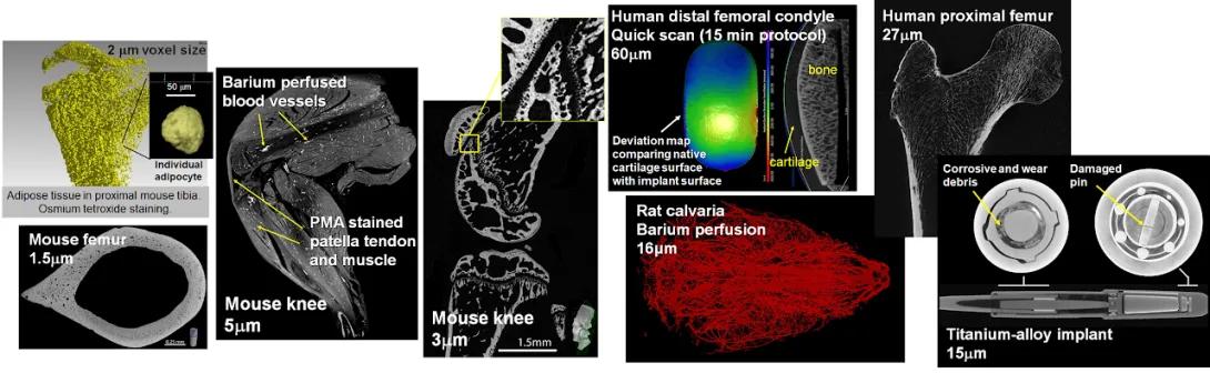
Composite of micro-CT and imaging scans showing bone, vascular, and implant structures in mice, rats, and humans at various resolutions.
• Top left: 3D rendering of adipose tissue in the proximal mouse tibia with osmium tetroxide staining. Inset shows an individual adipocyte at 2 μm voxel size.
• Bottom left: Transverse micro-CT scan of a mouse femur with 1.5 μm resolution. Scale bar 0.25 mm.
• Center left: Image of mouse knee showing Barium-perfused blood vessels and PMA-stained patella tendon and muscle. Resolution: 5 μm.
• Center: Mouse knee micro-CT scan at 3 μm voxel size, with an inset highlighting detailed cartilage and bone interface. Scale bar 1.5 mm.
• Top center: Human distal femoral condyle scan using a quick 15-minute protocol at 60 μm resolution. Includes a deviation map comparing native cartilage surface with implant surface. Bone and cartilage are labeled.
• Top right: Human proximal femur imaged at 27 μm resolution.
• Center right: Rat calvaria with Barium-perfused blood vessels shown in red. Resolution: 16 μm.
• Bottom right: Titanium-alloy implant at 15 μm resolution, showing detailed internal components including a damaged pin and corrosive/wear debris.
Services include ex vivo micro-Computed Tomography (CT) and ex vivo nano-Computed Tomography (nano-CT), Raman spectroscopy, and Histology as the major components.
The ex vivo microComputed Tomography (microCT) and nanoCT systems provide full 3-D reconstructed images of hard and soft biological tissues, as well as non-biological materials. The Dental School core has an ex vivo Scanco 100 microCT and the Orthopaedic Research Laboratories ex vivo scanner is a GE Phoenix nanotom-m nanoCT. See the websites listed below for details of system capabilities.
Raman spectroscopy services will be provided by Dr. Gurjit Mandair, who is located in the School of Dentistry. We provide guidance on sample preparation (fresh or embedded), compositional analysis (soft/hard tissues), and training on our Raman microscope/fiber-optic probe instruments
Histology is an essential component of all in vivo musculoskeletal studies. This core will enhance existing research by centralizing histologic efforts to achieve high quality and reasonably priced histology for center investigators, and to ensure, in collaboration with the other cores, a cost-effective, efficient, integrated, hierarchical approach to the evaluation of tissue phenotypes. Procedures are posted on the website.
The Histology part of this core supports these following procedures: plastic processing/embedding/sectioning and staining, paraffin processing/embedding/sectioning and staining, and frozen sectioning. Discounts are provided primarily for plastic procedures as the prices for paraffin procedures are very competitive with other facilities on campus.
An advanced histology training program has been developed to provide for the safe and proficient use of histological equipment. In addition to the training, supervision of first-time use is provided to be sure that all safety measures are being followed and the equipment is being used in the proper manner.
Each training session also comes with future technical support, slide Q & A, staining advice, etc., as our goal is to have trainees not just know how to produce histology products, but how to do it well and to understand what they are doing on a scientific level. Trainees are encouraged to ask our Histologists anything about the process, suggested processing adaptations, how to better prepare samples the next time, etc. We also have a stock of control tissue that is open for anyone completing the training.
Referencing Core services in manuscripts
When referencing the use of the MiMHC Core services in your manuscripts, please use the following text:
Research reported in this publication was supported by the National Institute of Arthritis and Musculoskeletal and Skin Diseases of the National Institutes of Health under Award Number P30 AR069620. The content is solely the responsibility of the authors and does not necessarily represent the official views of the National Institutes of Health.
If utilizing the ORL Histology, ORL Nano-CT, or Dental School MicroCT cores, please include the appropriate Research Resource Identifiers (RRIDs) when citing the resource:
- University of Michigan ORL Histology Core RRID:SCR_026775
- University of Michigan ORL Nano-CT Core RRID:SCR_026772
- University of Michigan Dental School MicroCT Core RRID:SCR_026747
Contact
Contact David Kohn, PhD when beginning a project and for general assistance with core access. He will provide guidance on experimental design and the appropriate Faculty and Core expert to contact next.