Functional Assessment Core
Bringing together animal and organ level phenotyping resources
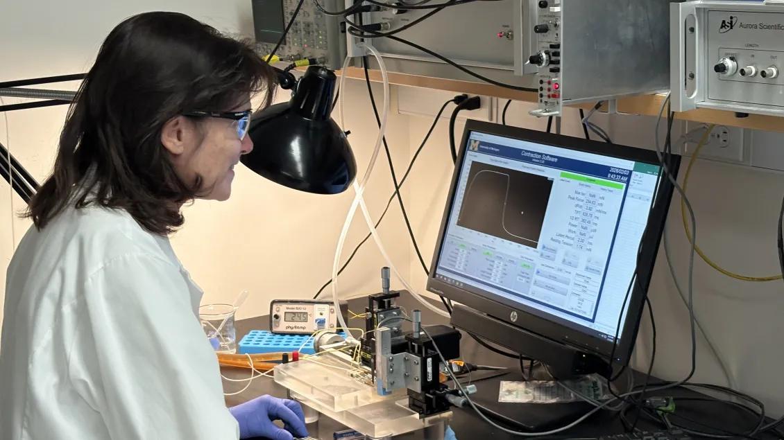
The overall long term goal of the Functional Assessment Core is to bring together the animal and organ level phenotyping resources currently used within individual laboratories and departmental cores at the University of Michigan into a cohesive and easily accessible Core service that can be used by the entire musculoskeletal research community. This core will provide access to state of the art whole animal phenotyping, muscle function and mechanical testing from fibers to whole muscles in situ, tendon and ligament mechanical testing, whole bone mechanical testing, and tissue-level bone mechanical testing. This core will enable center investigators to establish the functional context of animal models to targeted genetic or pharmacological perturbations. Further, the broad technology base and solid communication plan and expert guidance will allow center investigators to expand on their tissue of interest to also include assays on adjacent tissues to test for effects across tissues.
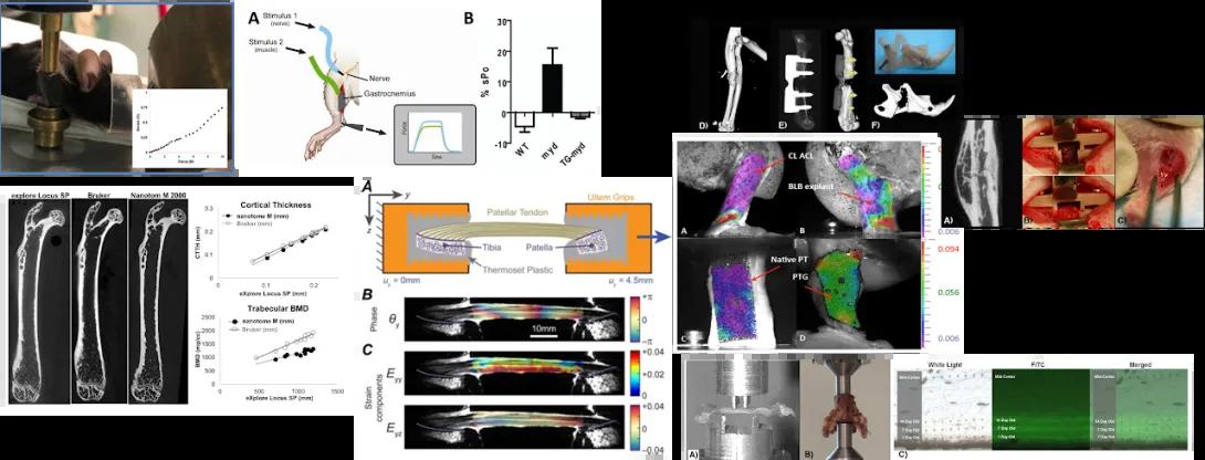
Composite figure showing various biomechanical and imaging analyses of musculoskeletal tissues, surgical procedures, and material testing, likely in the context of orthopedic or tendon research.
⸻
Top Left Section:
• Image of an animal with the leg clamped in a mechanical device for force testing.
• Inset: Graph showing increased muscle force or tension in response to stimulation.
• Diagram of a sciatic nerve stimulation setup with two electrode sites, a nerve, and the gastrocnemius muscle.
• Bar graph (Panel B) showing percentage force difference across genotypes: WT, MT1, and Transplanted.
⸻
Middle Left Section:
• Micro-CT scans of long bones from various mouse genotypes (e.g., WT, KO), showing differences in cortical thickness and trabecular bone.
• Quantitative graphs of cortical thickness, bone mineral density (BMD), and structure based on genotype.
⸻
Center Section (Orange background):
• Schematic of a mechanical testing device showing tensile loading across a patellar tendon-to-bone interface using Ultem grips and a thermoset plastic stage.
• Heatmap images of phase and strain fields across the tendon-bone interface under mechanical loading. Panels B and C show displacement and strain across the interface.
⸻
Top Right Section:
• 3D reconstructions of a limb and joint replacement components from micro-CT scans (Panels D–F).
• Multicolor strain mapping overlays on joint explants and native patellar tendon graft (PTG), highlighting load transfer and strain gradients.
• Panels labeled A–D show views of strain field comparisons between control ligaments (CL), anterior cruciate ligament (ACL), and bone-ligament-bone (BLB) explants.
⸻
Far Right Column:
• Micro-CT slice showing bone integration.
• Two surgical photographs showing orthopedic procedures with fixation pins and surgical manipulation.
⸻
Bottom Row:
• Mechanical testing of bone or tissue samples using a compression/tensile load frame (Panels A and B).
• Fluorescent imaging of tissue sections under white light and FITC channel (Panel C), with columns labeled “White Light,” “FITC,” and “Merged,” likely showing tagged markers or integration of labeled material.
The Core brings together the leadership and technical proficiency in the physiology and biomechanics of the musculoskeletal system along with specialized laboratory approaches to musculoskeletal biology including state-of-the-art measurements in the following areas:
- Bone composition and mechanics
- Muscle function and mechanics
- Tendon and soft tissue mechanics
- Neuromuscular function at the whole animal level
- The adaptations and regeneration of these tissues in response to injury and disease
Referencing Core services in manuscripts
When referencing the use of the MiMHC Core services in your manuscripts, please use the following text:
Research reported in this publication was supported by the National Institute of Arthritis and Musculoskeletal and Skin Diseases of the National Institutes of Health under Award Number P30 AR069620. The content is solely the responsibility of the authors and does not necessarily represent the official views of the National Institutes of Health.
Contact
Contact Dan Michele, PhD (whole animal testing, micro-surgery models), Ken Kozloff, PhD (bone testing, fracture healing/surgical models, in vivo microCT imaging), Ellen Arruda, PhD (tendon testing), or Susan Brooks, PhD (muscle mechanics) when beginning a project. Contact Dr. Michele, Dr. Kozloff, or Dr. Arruda if you are unsure whom to contact. They will provide guidance on experimental design and the appropriate Faculty and/or Core expert to contact next.