Protective Mechanisms & Pathways
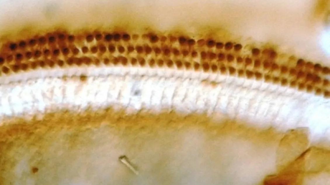
Induction of Heat Shock Protein 70 in outer hair cells following noise
Model of stress pathways in the cochlea
We study the stress response in the cochlea and auditory brain stem, specifically the pathways that lead to protection, repair and recovery. The genes and proteins within these pathways as well as their interaction and regulation are identified. Interventions are being developed, based on these pathways, to improve the efficacy of protection, repair and recovery and ultimately reduce acquired deafness.
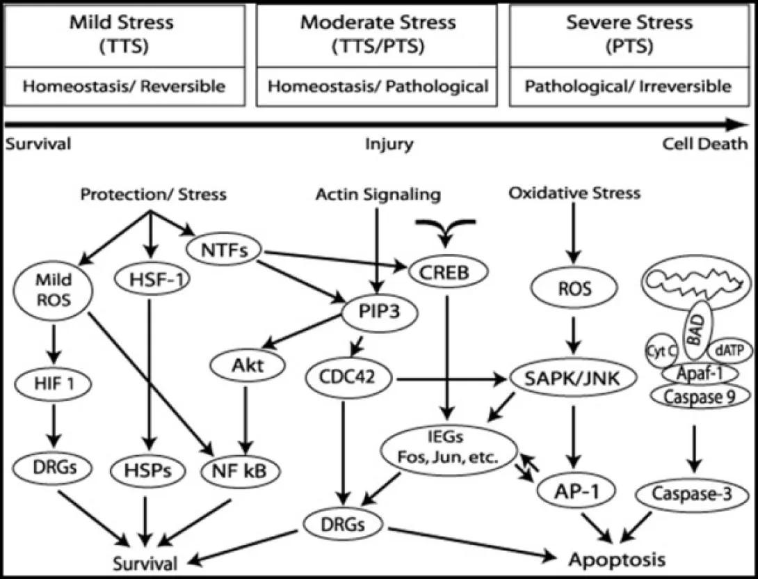
Diagram of cellular stress responses, divided into three columns labeled “Mild Stress (TTS) – Homeostasis/Reversible,” “Moderate Stress (TTS/PTS) – Homeostasis/Pathological,” and “Severe Stress (PTS) – Pathological/Irreversible.” Below these headers runs a horizontal arrow from “Survival” at the left, through “Injury” in the middle, to “Cell Death” at the right.
• Under Mild Stress (left): A “Protection/Stress” arrow splits to three ovals: Mild ROS, HSF-1, and NTFs.
• Mild ROS → HIF-1 → DRGs → survival.
• Mild ROS → HSF-1 → HSPs → survival.
• HSF-1 and Mild ROS both activate NF-κB → survival.
• Under Moderate Stress (center):
• “Actin Signaling” and NTFs both feed into PIP3.
• PIP3 activates Akt and CDC42, both of which promote DRGs.
• PIP3 and CDC42 also activate CREB, which together drive IEGs (Fos, Jun, etc.).
• IEGs link back to DRGs (toward survival) or to SAPK/JNK and AP-1 (toward apoptosis/injury).
• Under Severe Stress (right):
• Oxidative Stress → ROS → SAPK/JNK → AP-1 → apoptosis.
• ROS triggers mitochondrial release of cytochrome c, BAD, and dATP → Apaf-1/Caspase 9 → Caspase 3 → apoptosis.
Overall, mild stress pathways converge on protective stress proteins and gene programs for survival; moderate stress engages mixed signaling (PIP3/CREB/IEGs) that can lead either to adaptive gene expression (DRGs) or to stress-activated kinases; severe stress tips the balance toward SAPK/JNK signaling and mitochondrial caspase activation, driving irreversible cell death.
Heat Shock Proteins
We are examining the pathway involving heat shock proteins and their protective function in the auditory pathways. We study the expression of heat shock proteins such as Hsp70, Hsp110 and Hsp32 induced by heat, noise or ototoxins as well as constitutive heat shock proteins such as Hsp27 and their role following stress. We are also studying the role of Heat Shock Factors (Hsf), which regulate the production of heat shock proteins and other stress response genes. We find expression of Hsf1 in the cochlea and its activation by stress. We find that the conditions which induce heat shock proteins provide protection from a subsequent noise exposure (during the time that hearing has recovered but HSPs are still elevated), that would normally be damaging and that there is less recovery from noise in mice in which the Hsf1 gene has been knocked out. We also find that in mice with the Hsf1 gene knocked-out, that there is reduced recovery from noise overstimulation.
Heat Shock Factor 1 in the rat and mouse cochlea
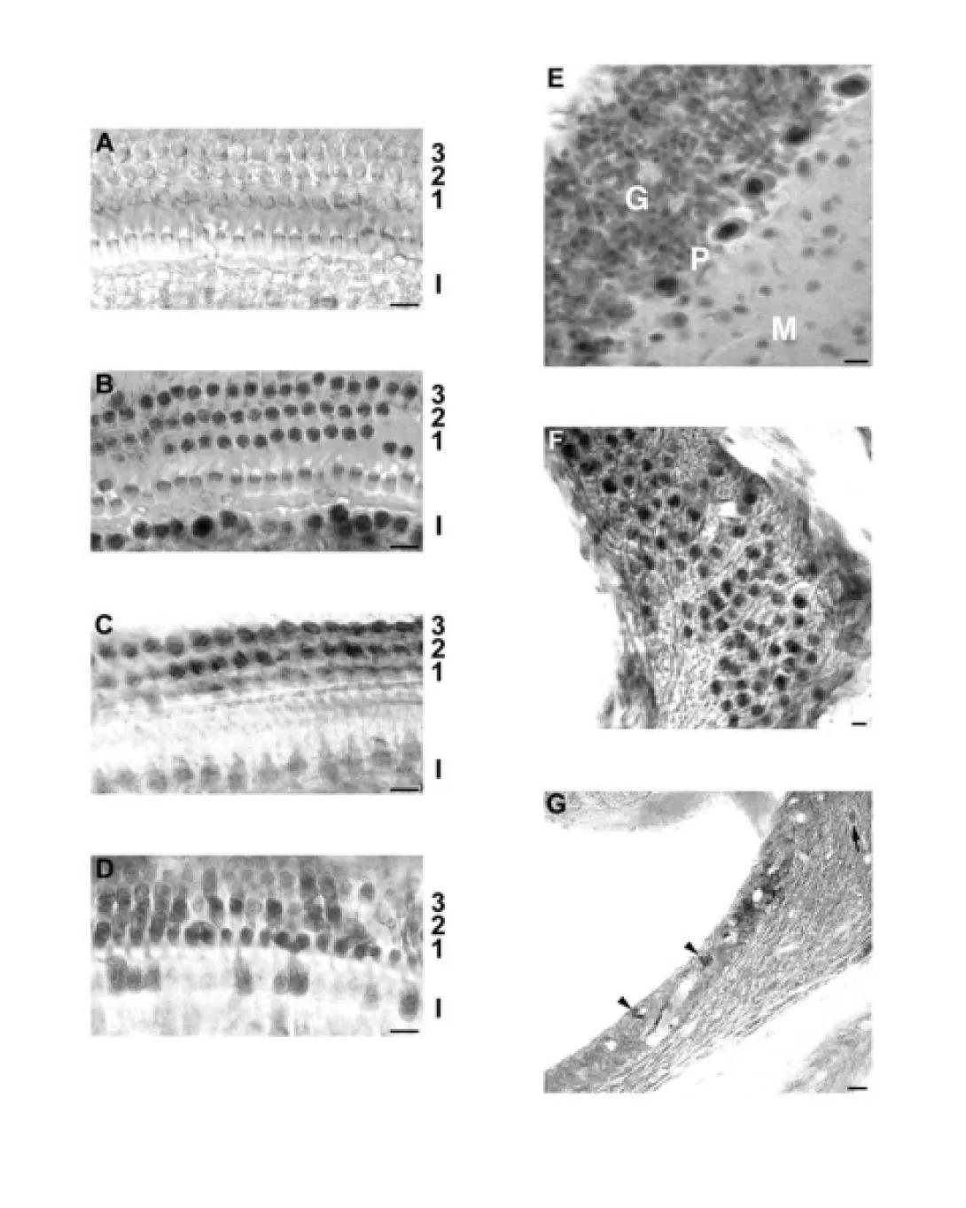
A composite of grayscale micrographs showing expression of Heat Shock Factor 1 (HSF1) in the cochlea of rats and mice.
• Panels A–D: Cross-sections of the organ of Corti showing immunolabeling of HSF1 in three rows of outer hair cells (labeled 3, 2, 1 from top to bottom) and the inner hair cell region (labeled I).
• Panel A: Control or baseline condition showing light, diffuse labeling.
• Panel B–D: Increased nuclear staining in outer and inner hair cells, likely under stress conditions or after heat shock.
• Panel E: Section through the cochlear ganglion. Labeled regions: G (ganglion cells), P (perineuronal area), and M (modiolus), showing distinct nuclear labeling in ganglion cells.
• Panel F: Section through cochlear nerve fibers with strong nuclear staining in supporting cells.
• Panel G: Peripheral cochlear nerve region with arrowheads pointing to nuclei with strong HSF1 expression.
All scale bars represent ~10 microns. The images demonstrate that HSF1 is present and activated in cochlear sensory and neural structures under stress, suggesting a role in protective stress responses.
Mice with a "knock-out" of Hsf1 exhibit decreased recovery from noise
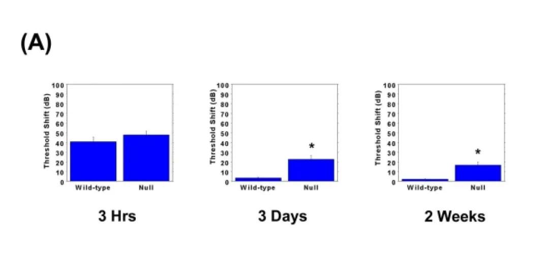
Bar graph labeled (A) showing auditory threshold shift (in decibels) over three time points—3 Hours, 3 Days, and 2 Weeks—comparing Wild-type and Null (HSF1 knockout) mice following noise exposure.
• Y-axis: Threshold Shift (dB), ranging from 0 to 100 dB.
• X-axis: Time post-exposure:
• 3 Hrs: Wild-type and Null mice show similar elevated threshold shifts (~50 dB).
• 3 Days: Wild-type mice recover (near 0 dB shift), while Null mice show a residual shift (~30 dB). An asterisk (*) indicates statistical significance.
• 2 Weeks: Wild-type mice maintain recovery, Null mice retain a moderate threshold shift (~25 dB), again marked with an asterisk for significance.
Interpretation: Null mice lacking HSF1 show impaired recovery from noise-induced hearing loss compared to wild-type mice.