Publications | Hammoud Lab
Explore Our Latest Publications
Dive into our extensive collection of research publications covering a wide range of topics and disciplines.
Featured Publications
ZCWPW1 organizes telomeric architecture to drive meiotic chromosome movements
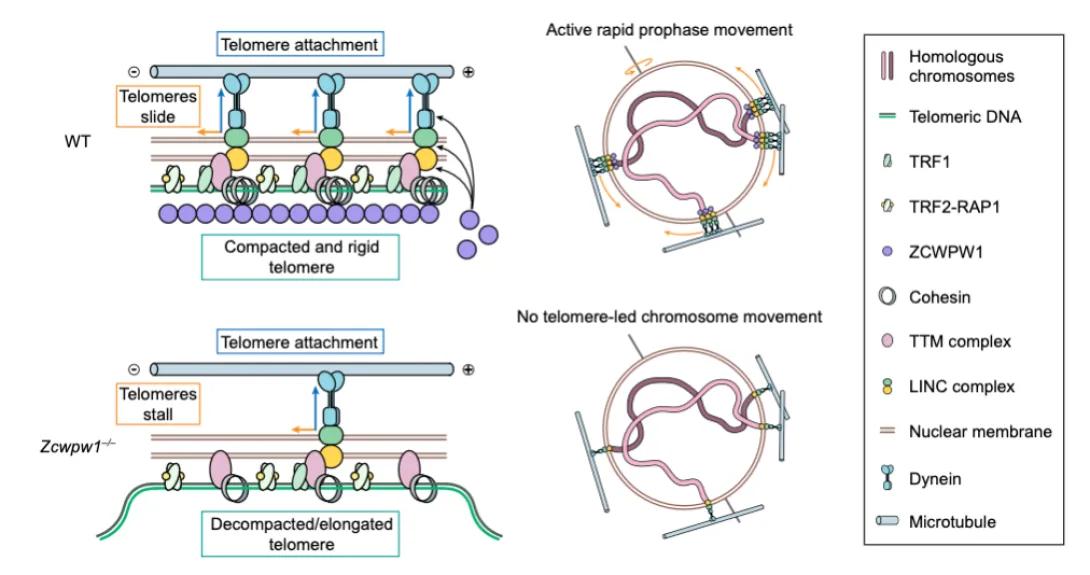
Schematic diagram comparing telomere structure and chromosome movement in wild-type versus Zcwpw1-deficient mouse spermatocytes during meiotic prophase. In wild-type cells (top), compact telomeres attach to the nuclear envelope via the LINC complex and dynein, slide along microtubules, and drive active rapid prophase chromosome movements. In Zcwpw1−/− cells (bottom), telomeres are decompacted and elongated, fail to properly couple to the LINC–motor machinery, and stall, resulting in no telomere-led chromosome movement. A legend identifies molecular components including telomeric DNA, TRF proteins, ZCWPW1, cohesin, the LINC complex, dynein, microtubules, and the nuclear membrane.
ZCWPW1 is required for telomere-led chromosome movements during meiotic prophase, acting independently of its known role as a PRDM9-associated histone reader. It localizes to subtelomeric regions to stabilize telomere architecture and the telomere–LINC–motor axis; loss of ZCWPW1 abolishes chromosome movement, disrupts synapsis, and leads to persistent DNA breaks.
A hidden protamine PTM code in sperm generates heterogeneous chromatin states and finetunes reproductive fitness
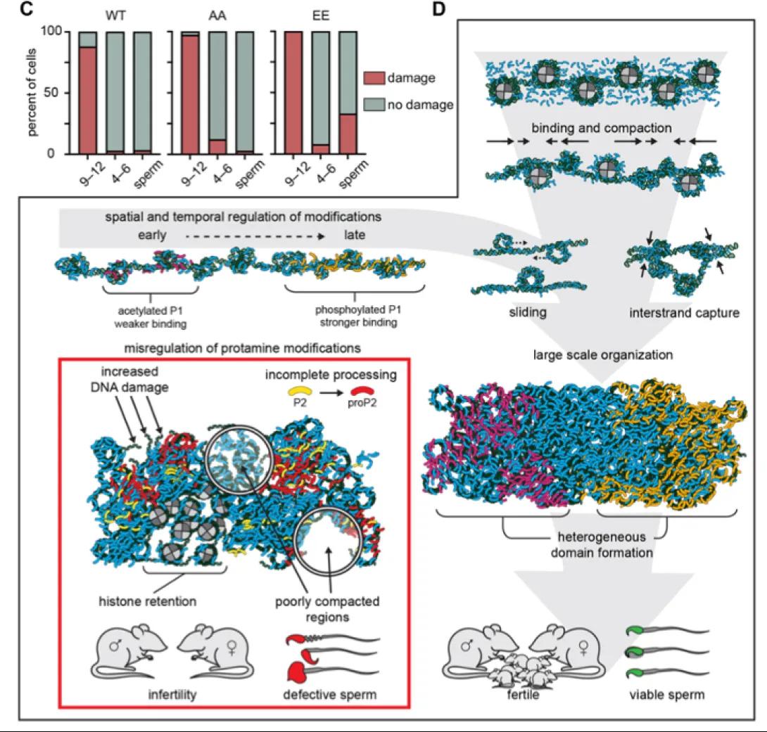
Multi-panel schematic illustrating how protamine post-translational modifications regulate sperm chromatin organization and fertility. A bar graph (panel C) compares DNA damage across developmental stages in wild-type and mutant sperm, showing increased damage when protamine modifications are altered. Diagrams depict a normal progression from weakly bound, acetylated protamine–DNA interactions to more strongly bound, phosphorylated states that enable binding, sliding, interstrand capture, large-scale chromatin organization, and formation of heterogeneous chromatin domains, producing viable sperm. In contrast, misregulation of protamine modifications leads to incomplete protamine processing, histone retention, poor compaction, elevated DNA damage, defective sperm, and infertility.
This study overturns the view of sperm chromatin as uniformly compact by showing that protamine post-translational modifications generate distinct protamine–DNA states with varying binding strength. Loss of these modifications disrupts sperm chromatin integrity and fertility, revealing that protamine PTMs create functional chromatin heterogeneity and likely encode instructions for paternal genome reorganization after fertilization.
Maternal CENP-C restores centromere symmetry in mammalian zygotes to ensure proper chromosome segregation
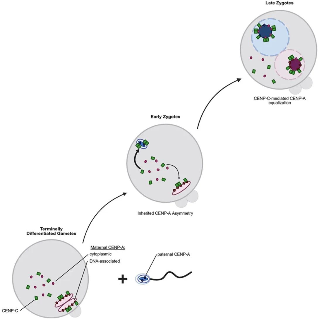
Diagram illustrating how centromeric CENP-A levels are equalized after fertilization. Terminally differentiated gametes contribute asymmetric centromeres, with sperm containing low levels of DNA-associated CENP-A and oocytes providing abundant maternal CENP-A and CENP-C, including a cytoplasmic pool. In early zygotes, this asymmetry is retained between paternal and maternal pronuclei. In late zygotes, maternally supplied CENP-C is recruited to paternal centromeres, promoting CENP-A incorporation and equalization between parental genomes prior to the first mitosis.
This study reveals that although mature sperm retain far less CENP-A than oocytes, centromeric CENP-A levels are equalized in the zygote before the first mitosis using maternally inherited CENP-A. This process depends on asymmetric recruitment of maternal CENP-C to paternal centromeres, identifying maternal CENP-C as a key epigenetic regulator that restores centromeric symmetry and ensures faithful chromosome segregation after fertilization.
Distinctive chromatin in human sperm packages genes for embryo development
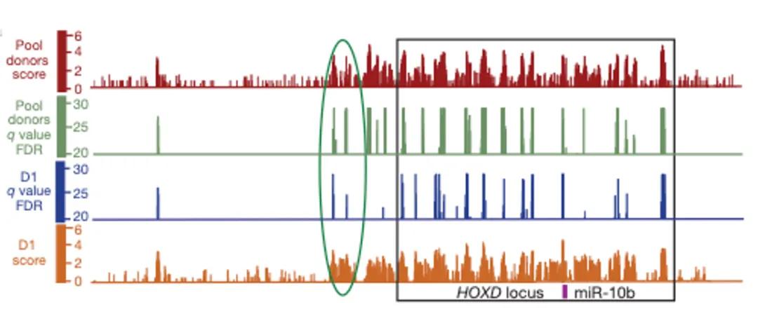
A multi-track genomic signal plot showing data across a genomic region labeled the HOXD locus, with miR-10b marked near the right side. Four horizontal tracks are stacked vertically. From top to bottom: (1) red bars representing “Pool donors score,” (2) green vertical spikes representing “Pool donors q value (FDR),” (3) blue vertical spikes representing “D1 q value (FDR),” and (4) orange bars representing “D1 score.” The x-axis represents genomic position, while the y-axes show signal intensity or statistical significance for each track. A green oval highlights a specific region where signals increase across multiple tracks. A black rectangular box outlines a broader region of interest spanning the HOXD locus. The highlighted areas emphasize regions with elevated scores and significant q values across datasets.
Although most nucleosomes are replaced by protamines in human sperm, this study shows that retained nucleosomes are selectively enriched at key developmental loci, including imprinted genes, HOX clusters, and developmental promoters. These regions carry distinct histone modifications and low DNA methylation, demonstrating that sperm chromatin retains extensive epigenetic information correlated with embryonic gene regulation.
A Comprehensive Roadmap of Murine Spermatogenesis Defined by Single-Cell RNA-Seq
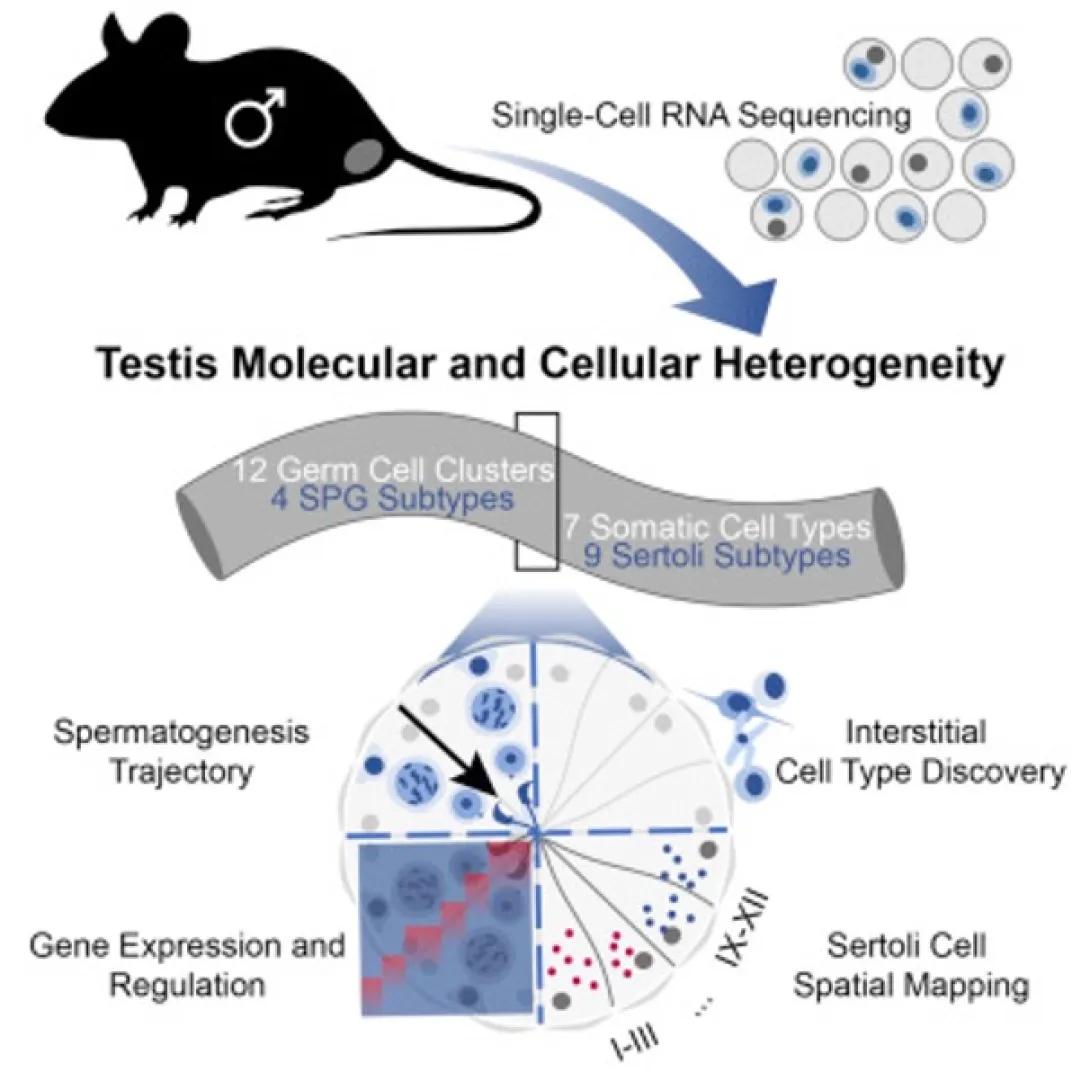
A schematic infographic illustrating a study of testis molecular and cellular heterogeneity using single-cell RNA sequencing. At the top left, a silhouette of a male mouse is shown, with an arrow pointing to a cluster of individual cells, indicating single-cell RNA sequencing. The central title reads “Testis Molecular and Cellular Heterogeneity.” Below, a curved banner summarizes results, stating the identification of 12 germ cell clusters including 4 spermatogonia (SPG) subtypes, and 7 somatic cell types including 9 Sertoli cell subtypes. The lower portion of the image shows a circular diagram representing spermatogenesis progression, divided into stages labeled I–III and IX–XII. Within the diagram are illustrated cells with varying gene expression patterns. Additional labels indicate key analyses: spermatogenesis trajectory, gene expression and regulation, interstitial cell type discovery, and Sertoli cell spatial mapping. The figure visually summarizes how single-cell sequencing reveals diverse cell types and developmental pathways in the testis.
Single-cell RNA sequencing of ~35,000 adult mouse testis cells resolves the cellular heterogeneity of spermatogenesis, identifying all known germ and somatic cell types plus two previously unrecognized somatic populations. This high-resolution atlas reveals continuous germ cell differentiation, distinct spermatogonia and Sertoli cell subtypes, and key transcriptional regulators, providing a foundational resource for studying in vivo gametogenesis.
Single-cell RNA sequencing of human, macaque, & mouse testes uncovers conserved & divergent feat. of mammalian spermatogenesis
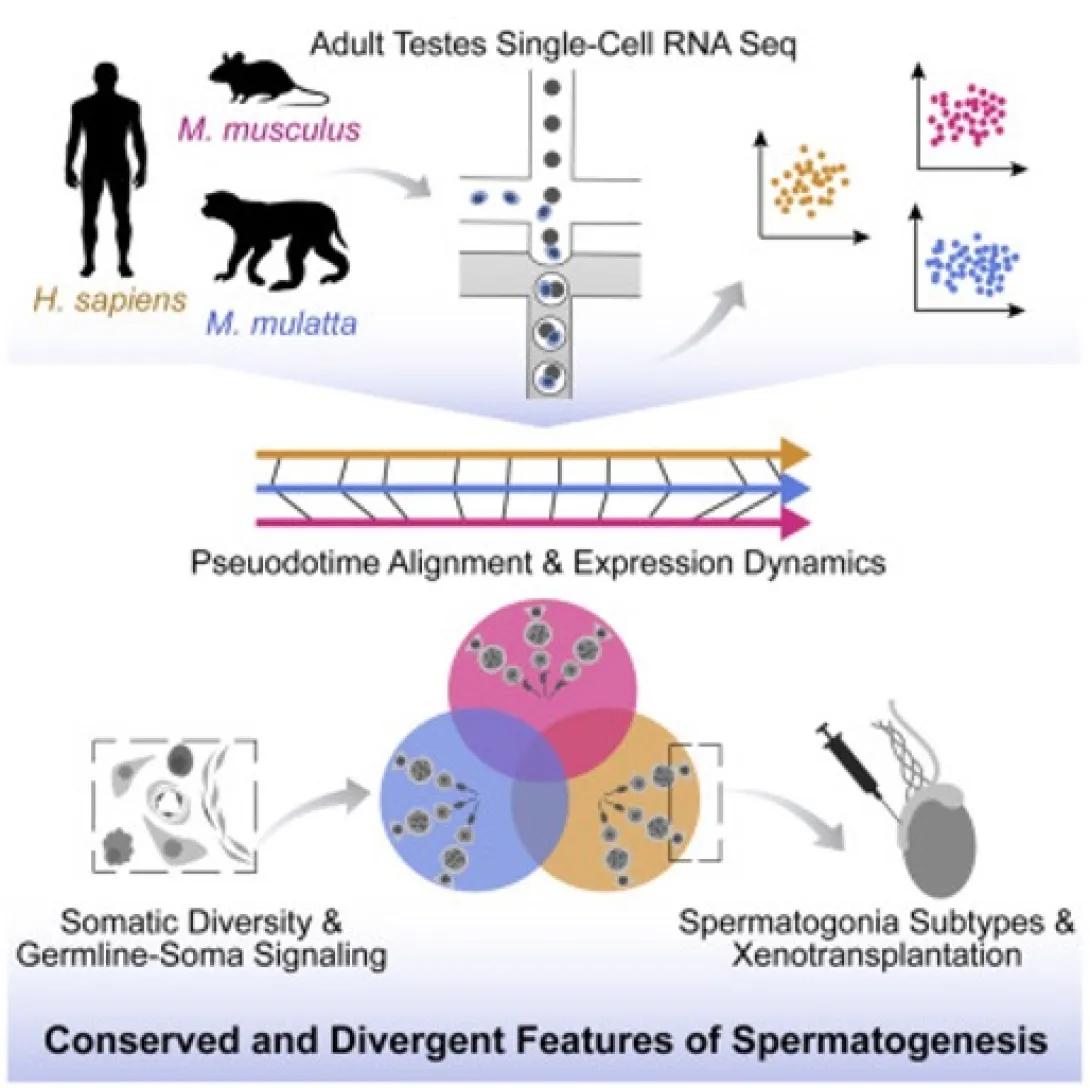
An infographic summarizing a comparative single-cell RNA sequencing study of adult testes across species. At the top, silhouettes of a human (Homo sapiens), mouse (Mus musculus), and macaque (Macaca mulatta) indicate cross-species analysis. These are connected to a schematic of single-cell RNA sequencing, which branches into colored scatter plots representing distinct cell populations. The middle section shows a horizontal timeline labeled “Pseudotime Alignment & Expression Dynamics,” illustrating the alignment of gene expression changes during spermatogenesis across species. The lower section includes three overlapping circles representing integrated analyses of cell populations, alongside icons depicting somatic cell diversity and germline–somatic signaling, as well as spermatogonia subtypes and xenotransplantation experiments. A caption at the bottom reads “Conserved and Divergent Features of Spermatogenesis,” highlighting similarities and differences in spermatogenic processes across species.
We used single-cell RNA sequencing to map transcriptional signatures of germ and somatic testis cells in humans, macaques, and mice, revealing both conserved and species-specific features of spermatogenesis. These datasets highlight differences in spermatogonial stem/progenitor pools, differentiation markers, meiotic regulators, and germ cell–somatic interactions, providing a foundation for future studies of primate germ cell development and in vitro gametogenesis.
TCF21+ mesenchymal cells contribute to testis somatic cell development, homeostasis, and regeneration in mice
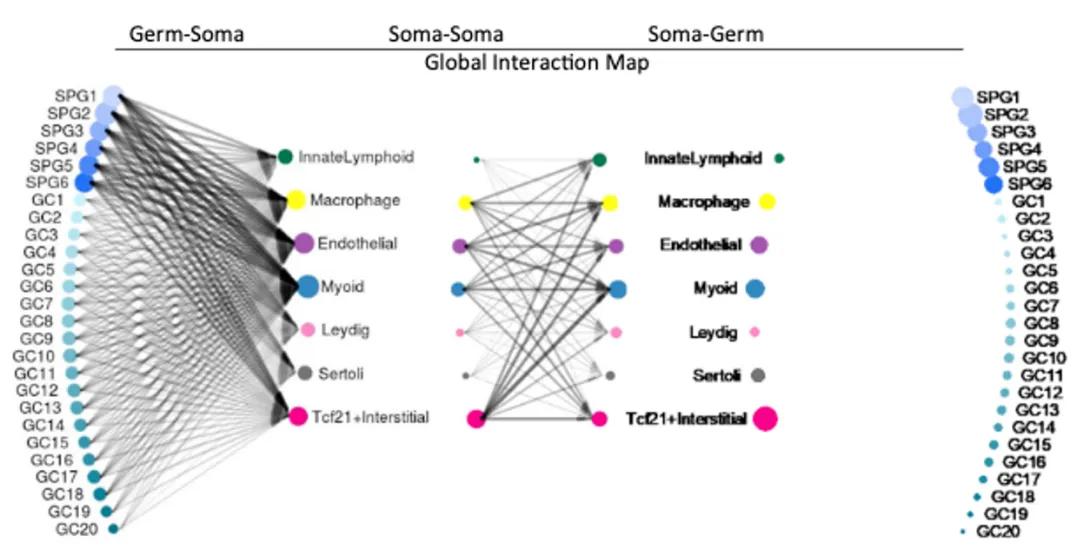
Network-style diagram showing a global interaction map between germ cells and somatic cell types in the testis. Germ cell populations (SPG1–SPG6 spermatogonia and GC1–GC20 differentiating germ cells) are displayed on the left and right, while somatic cell types—including Sertoli, Leydig, endothelial, macrophage, myeloid, innate lymphoid, and Tcf21+ interstitial cells—are shown in the center. Lines connecting nodes represent inferred cell–cell interactions, with dense connections illustrating extensive signaling between germ and somatic compartments, as well as interactions among somatic cell types themselves.
We characterize TCF21-expressing somatic progenitors in the mouse testis, showing they maintain homeostasis during aging, act as reserve progenitors after injury, and can differentiate into multiple somatic lineages in vitro. These findings reveal that the adult testis retains multipotent mesenchymal progenitors similar to fibroblast populations in other organs, with potential relevance for therapies targeting hypoandrogenism and infertility.
Cellular heterogeneity of human fallopian tubes in normal and hydrosalpinx disease states identified using scRNA-seq
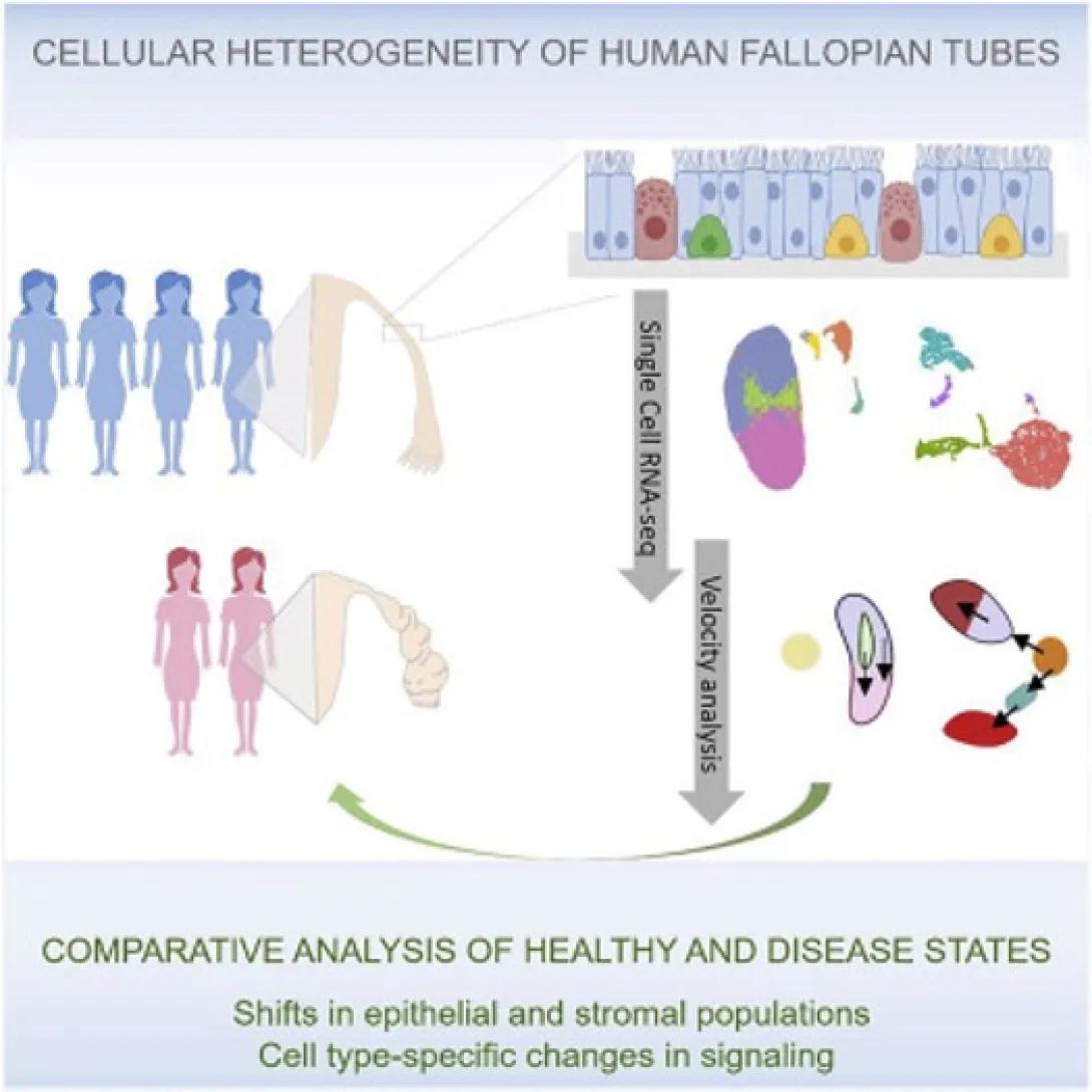
Schematic overview of a study analyzing cellular heterogeneity in human fallopian tubes. Illustrations of healthy and disease-affected individuals lead to a diagram of fallopian tube tissue, highlighting diverse epithelial cell types lining the tube. Single-cell RNA sequencing and computational analyses are shown identifying distinct cell populations, transcriptional states, and developmental trajectories. The figure emphasizes comparative analysis between healthy and diseased tissues, revealing shifts in epithelial and stromal cell populations and cell type–specific signaling changes.
We used single-cell RNA-seq to profile 59,738 fallopian tube cells, identifying 12 major cell types and epithelial subtypes, including two potential progenitor pools. Comparison with hydrosalpinx samples revealed disease-associated shifts in epithelial and stromal populations and signaling pathways, providing a resource for studying fallopian tube homeostasis and pathology.
Additional Publications
- Kelly M. Bakulski*, John F. Dou, Robert C. Thompson, Christopher Lee, Lauren Y. Middleton, Bambarendage P. U. Perera, Sean P. Ferris, Tamara R. Jones, Kari Neier, Xiang Zhou, Maureen A. Sartor, Saher S. Hammoud, Dana C. Dolinoy, Justin A. Colacino*. Single cell analysis of the gene expression effects of developmental lead (Pb) exposure on the mouse hippocampus. Toxicol Sci. epub 2020 May 27.
- Hailey Larose, Travis Kent, Qianyi Ma, Adrienne Niederriter Shami, Nadia Harerimana, Jun Z. Li, Saher Sue Hammoud*, Mary Ann Handel*. Regulation of meiotic progression by Sertoli-cell androgen signaling. bioRxiv preprint doi:https://doi.org/10.1101/2020.05.26.117093.
- Yu-chi Shen*, Hailey Larose*, Adrienne Niederriter Shami*, Lindsay Moritz*, Gabriel L. Manske#, Qianyi Ma#, Xianing Zheng, Meena Sukhwani, Michael Czerwinski, Caleb Sultan, Jourdan Clements, Haolin Chen, Jason R. Spence, Kyle E. Orwig, Michelle Tallquist, Jun Z. Li, Saher Sue Hammoud. Tcf21+ mesenchymal cells contribute to testis somatic cell development, homeostasis, and regeneration. bioRxiv preprint doi: https://doi.org/10.1101/2020.05.02.074518.
- Adrienne Niederriter Shami1,6, Xianing Zheng1,6, Sarah K. Munyoki2,6, Qianyi Ma1, Christopher D. Green1, Meena Sukhwani2, Kyle E. Orwig2, Jun Z. Li1,3, Saher Sue Hammoud1,4,5,7*. Single-cell RNA sequencing of human, macaque, and mouse testes uncovers conserved and divergent features of mammalian spermatogenesis. Dev Cell. epub. 2020 PMID: 32504559.
- Sohni A, Tan K, Song HW, Burow D, de Rooij DG, Laurent L, Hsieh TC, Rabah R, Hammoud SS, Vicini E, Wilkinson MF: The Neonatal and Adult Human Testis Defined at the Single-Cell Level. Cell Rep 26(6): 1501-1517.e4, 2019. PM30726734
- Green CD, Ma Q, Manske GL, Shami AN, Zheng X, Marini S, Moritz L, Sultan C, Gurczynski SJ, Moore BB, Tallquist MD, Li JZ, Hammoud SS: A Comprehensive Roadmap of Murine Spermatogenesis Defined by Single-Cell RNA-Seq. Dev Cell 46(5): 651-667.e10, 2018. PM30146481
- Dehghanizadeh S, Khoddami V, Mosbruger TL, Hammoud SS, Edes K, Berry TS, Done M, Samowitz WS, DiSario JA, Luba DG, Burt RW, Jones DA: Active BRAF-V600E is the key player in generation of a sessile serrated polyp-specific DNA methylation profile. PLoS One 13(3): e0192499, 2018. PM29590112 /PMC5873940
- Borges BC, Garcia-Galiano D, da Silveira Cruz-Machado S, Han X, Gavrilina GB, Saunders TL, Auchus RJ, Hammoud SS, Smith GD, Elias CF: Obesity-Induced Infertility in Male Mice Is Associated With Disruption of Crisp4 Expression and Sperm Fertilization Capacity. Endocrinology 158(9): 2930-2943, 2017. PM28911169
- Hammoud SS, Low DH, Yi C, Lee CL, Oatley JM, Payne CJ, Carrell DT, Guccione E, Cairns BR: Transcription and imprinting dynamics in developing postnatal male germline stem cells. Genes Dev 29 (21): 2312-24, 2015. PM26545815/PMC4647563
- Hammoud SS, Low DH, Yi C, Carrell DT, Guccione E, Cairns BR: Chromatin and transcription transitions of mammalian adult germline stem cells and spermatogenesis. Cell Stem Cell 15(2): 239-253, 2014. PM24835570
- Hong C, Clement NL, Clement S, Hammoud SS, Carrell DT, Cairns BR, Snell Q, Clement MJ, Johnson WE: Probabilistic alignment leads to improved accuracy and read coverage for bisulfite sequencing data. BMC Bioinformatics 14(1): 2013. PM24261665
- Hammoud SS, Cairns BR, Jones DA: Epigenetic regulation of colon cancer and intestinal stem cells Curr. Opin. Cell Biol. 25(2): 177-183, 2013. PM23402869
- Hammoud SS, Cairns BR, Carrell DT: Analysis of gene-specific and genome-wide sperm DNA methylation Methods Mol. Biol. 927: 451-458, 2013. PM22992936
- Swierczek SI, Piterkova L, Jelinek J, Agarwal N, Hammoud S, Wilson A, Hickman K, Parker CJ, Cairns BR, Cairns B, Prchal JT: Methylation of AR locus does not always reflect X chromosome inactivation state. Blood 119(13): e100-e109, 2012. PM22286197
- Wu SF, Zhang H, Hammoud SS, Potok M, Nix DA, Jones DA, Cairns BR: DNA methylation profiling in zebrafish. Methods Cell Biol. 104: 327-339, 2011. PM21924171
- Hammoud SS, Nix DA, Hammoud AO, Gibson M, Cairns BR, Carrell DT: Genome-wide analysis identifies changes in histone retention and epigenetic modifications at developmental and imprinted gene loci in the sperm of infertile men Hum. Reprod. 26(9): 2558-2569, 2011. PM21685136
- Milroy C, Liu L, Hammoud S, Hammoud A, Peterson CM, Carrell DT: Differential methylation of pluripotency gene promoters in in vitro matured and vitrified, in vivo-matured mouse oocytes. Fertil. Steril. 95(6): 2094-2099, 2011. PM21457962
- Hammoud SS, Purwar J, Pflueger C, Cairns BR, Carrell DT: Alterations in sperm DNA methylation patterns at imprinted loci in two classes of infertility. Fertil. Steril. 94(5): 1728-1733, 2010. PM19880108
- Carrell DT, Hammoud SS: The human sperm epigenome and its potential role in embryonic development. Mol. Hum. Reprod. 16(1): 37-47, 2009. PM19906823
- Hammoud SS, Nix DA, Zhang H, Purwar J, Carrell DT, Cairns BR: Distinctive chromatin in human sperm packages genes for embryo development. Nature 460(7254): 473-478, 2009. PM19525931
- Hammoud S, Emery BR, Dunn D, Weiss RB, Carrell DT: Sequence alterations in the YBX2 gene are associated with male factor infertility. Fertil. Steril. 91(4): 1090-1095, 2009. PM18339382
- Hammoud S, Liu L, Carrell DT: Protamine ratio and the level of histone retention in sperm selected from a density gradient preparation. Andrologia 41(2): 88-94, 2009. PM19260844
- Hammoud S, Emery BR, Aoki VW, Carrell DT: Identification of genetic variation in the 5′ and 3′ non- coding regions of the protamine genes in patients with protamine deregulation. Arch. Androl. 53(5): 267- 274, 2008. PM18309899
- Carrell DT, Emery BR, Hammoud S: The aetiology of sperm protamine abnormalities and their potential impact on the sperm epigenome. Int. J. Androl. 31(6): 537-545, 2008. PM18298569
- Carrell DT, Emery BR, Hammoud S: Altered protamine expression and diminished spermatogenesis: What is the link? Hum. Reprod. Update 13(3): 313-327, 2007. PM17208950