Positron Emission Tomography (PET) Imaging
The PET Center produces and develops non-invasive PET imaging techniques and radiopharmaceuticals to personalize medicine, advance our understanding of disease, and improve public health.
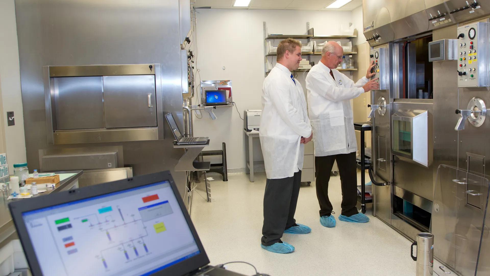
PET (positron emission tomography) is a non-invasive nuclear medicine imaging technique that uses drug-like molecules labeled with radioactive nuclides to elucidate biochemical processes, diagnose disease, and track the impact of experimental drugs in clinical trials. The PET Center conducts and supports PET imaging research studies at Michigan Medicine. The PET Radiochemistry group is focused on producing radiopharmaceuticals for PET imaging studies, and actively participates in industry-supported collaborative research projects in radiochemistry and preclinical studies of new drugs and radiopharmaceuticals. We routinely prepare radiopharmaceuticals for clinical application in cardiology, neurology and oncology using 11C, 18F, and 68Ga.
Explore Positron Emission Tomography (PET) Imaging
During a PET scan, the PET-CT scanner detects the energy signature (pairs of gamma rays at a specific energy) that is emitted by the radiopharmaceutical.
Images of radioactivity concentration within the body are then reconstructed with computer analysis. This reconstruction is typically accomplished with the aid of a low-dose CT scan performed on the patient during the same procedure. Analysis of how the images change over time (kinetic modeling) allow for measurements of drug kinetics in living people.
The PET Center actively participates in industry-supported collaborative research projects in radiochemistry and preclinical studies of new drugs and radiopharmaceuticals, and provides routine delivery of radiopharmaceuticals for clinical trials with partners in the pharmaceutical and radiopharmaceutical drug industry.
Funding
Funding for our research from the following sources is gratefully acknowledged:
University of Michigan Undergraduate Research Opportunity Program
Contact Us
General Inquiries
Please contact us for further details and recharge pricing at [email protected].