Research | Humphries Lab

Metabolic Cancer Drivers
Our research investigates how breast cancer cells adapt, survive and evolve by examining two fundamental drivers of tumor behavior: mitochondrial metabolism and interactions with the extracellular matrix. By uncovering how these systems shape tumor-initiating cell physiology, signaling heterogeneity and metabolic reprogramming, we aim to reveal mechanisms that fuel breast cancer initiation, progression and therapy resistance.
Mitochondria & Cellular Metabolism
Tumor-initiating cells (TICs, also known as cancer stem cells (CSCs)) are a major contributor to breast tumor heterogeneity, leading to breast cancer initiation, progression, metastasis, and recurrence. Our current understanding of mechanisms that establish and maintain TIC physiology is incompletely defined. However, we have previously shown that phosphatidylserine decarboxylase (PISD) is a novel regulator of TICs by regulating multiple facets of mitochondrial morphology, physiology, and cellular metabolism, underlining mitochondrial functions as critical drivers of TICs. Therefore, this project aims to understand mitochondria as central regulators of TIC and breast cancer physiology.
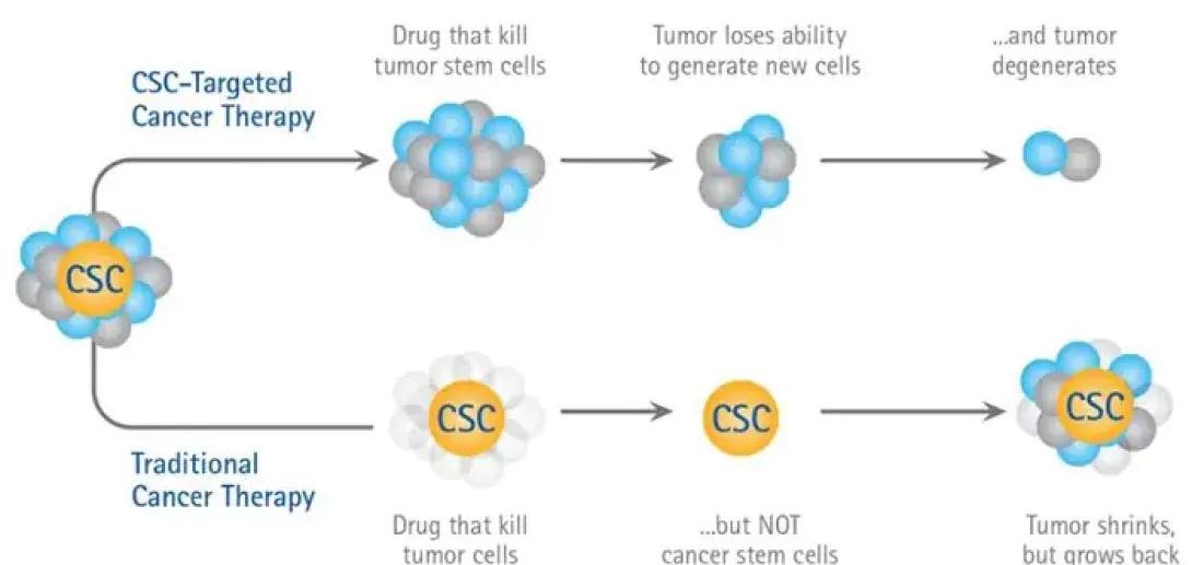
The graphic compares two approaches to cancer treatment: CSC-targeted therapy and traditional cancer therapy. At the top, a series of illustrations shows CSC-targeted therapy. It begins with a cluster of cells that includes a cancer stem cell (labeled “CSC” in a yellow center). An arrow points to a second cluster where a drug kills the tumor stem cells, shown as the blue and gray cells shrinking or disappearing. A third image shows a reduced cluster with fewer cells and a label explaining that the tumor loses its ability to generate new cells. A final image shows only two small remaining cells, illustrating that the tumor degenerates when cancer stem cells are eliminated.
Below, a parallel sequence represents traditional cancer therapy. The first image again shows a cluster with a CSC in the middle. A second image shows a drug that kills only tumor cells, not the cancer stem cell, leaving the CSC intact. The next image displays the single surviving CSC. A final panel shows the tumor growing back around the CSC, returning to a full cluster of cells. A caption notes that while the tumor initially shrinks, it eventually regrows because the cancer stem cell was not eliminated.
Interactions with the Extracellular Matrix
The behavior of cancer cells is directly influenced by the extracellular matrix (ECM), which provides both mechanical and biochemical cues to the cell. These cues activate intracellular signaling networks that drive metabolic reprogramming, survival, proliferation and motility. We have previously shown that biochemical cues, such as chemokines and chemotherapeutics, greatly influence key signaling molecules in breast cancer cells, contributing to overall cellular heterogeneity. Therefore, this project aims to understand ECM-dependent mechanisms linking signaling heterogeneity and metabolic reprogramming to differences in breast tumor initiation, progression, and treatment.
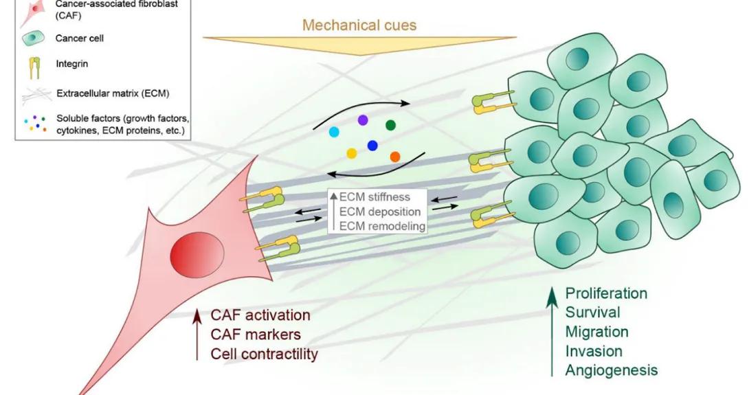
Diagram illustrating how mechanical cues in the tumor microenvironment influence cancer progression. On the left, a large pink cancer-associated fibroblast (CAF) connects to a network of extracellular matrix (ECM) fibers, shown as crisscrossing gray lines. The CAF displays increased activation, CAF markers and cell contractility. Soluble signaling factors (colored dots) circulate between the CAF and a cluster of green cancer cells on the right. Both the CAF and the cancer cells attach to the ECM through yellow integrins. A central box highlights ECM changes, including increased stiffness, ECM deposition and ECM remodeling. These mechanical and biochemical cues promote cancer cell behaviors such as proliferation, survival, migration, invasion and angiogenesis. A legend in the upper left identifies the symbols for CAFs, cancer cells, integrins, ECM and soluble factors.