Spatialomics
Information regarding tissue prep and sample submission for Visium, GeoMx DSP, Curio Seeker, Xenium, and Aviti24 CytoProfiling processing.
On this page...
10X Genomics Visium
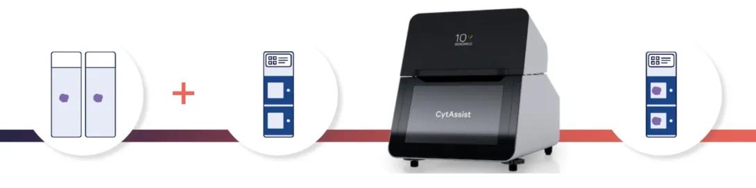
The Visium CytAssist workflow follows a standard histological workflow: sectioning, tissue preparation, staining (H&E or IF), and imaging all take place on a standard glass slide. Additional tissue preparation is followed by probe hybridization on the same glass slide. During this phase, probes hybridize to approximately 18,000 genes, or RNA targets, within the tissue section, offering whole transcriptome gene expression profiling.
Next, the CytAssist instrument incorporates two normal glass slides and a Visium slide with a pair of Capture Areas. This arrangement allows for the alignment of tissue sections on the standard slides with the Capture Areas.
The instrument uses a brightfield image for spatial referencing during data analysis, and then the transcriptomic probes from the tissue hybridize onto the Visium slide. The steps that follow, starting with probe extension, adhere to the standard Visium procedure outside of the instrument.
Visium HD (CytAssist)
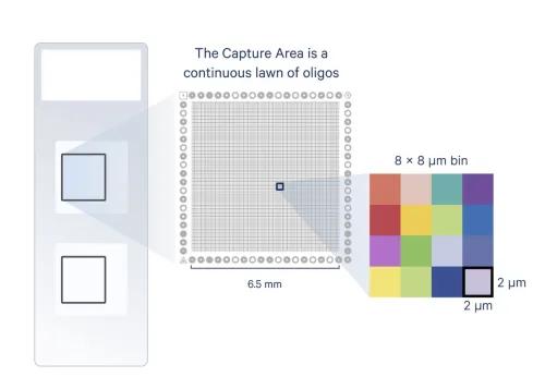
Visium HD uses the CytAssist workflow. HD slides contain two 6.5 x 6.5 mm or 11 x 11 mm Capture Areas with a continuous lawn of oligonucleotides, arrayed in millions of 2 x 2 µm barcoded squares without gaps, achieving single cell-scale spatial resolution. The data is output at 2 µm, as well as multiple bin sizes. The 8 x 8 µm bin is the recommended starting point for visualization and analysis.
CytAssist Workflow: Investigators section tissue onto standard glass slides. Once tissue is placed on the slide, the slide is transported to the AGC. AGC staff will perform H&E staining, imaging, library prep, sequencing, and SpaceRanger data processing.
Tissue Submission: Tissue processed following the recommended protocols can be submitted directly to the AGC for sectioning, staining/imaging, library prep, sequencing, and data processing. The cryomold used for embedding should be of appropriate size to fit the tissue sample. Preferred size for cryoblock submission is 10 mm.
Sample Requirements
- Species Specific: Only human, mouse, and rat samples are currently supported
- Fresh/Frozen, Fixed/Frozen, or FFPE
- Ensure desired tissue area will fit within the 6.5 x 6.5 mm or 11 x 11 mm capture area
- Section tissue within the allowable target area on the standard glass slide
- Recommended section thickness is 3-10 µm
- RNA quality of the block should be assessed prior to sectioning.
- recommended DV200 ≥ 30%
Visium HD 3' (CytAssist)
CytAssist Workflow: Investigators section tissue onto standard glass slides. Once tissue is placed on the slide, the slide is transported to the AGC. AGC staff will perform H&E staining, imaging, library prep, sequencing, and SpaceRanger data processing.
Tissue Submission: Tissue processed following the recommended protocols can be submitted directly to the AGC for sectioning, staining/imaging, library prep, sequencing, and data processing. The cryomold used for embedding should be of the appropriate size to fit the tissue sample. Preferred size for cryoblock submission is 10 mm.
Sample Requirements
- Freshly obtained tissue should be snap frozen to prevent RNA degradation
- The recommended freezing uses an isopentane and liquid nitrogen bath
- Frozen tissue should be embedded in Optimal Cutting Temperature (OCT), a freezing and embedding compound (can be done simultaneously with freezing process)
- RNA quality of the tissue block should be assessed prior to sectioning. The RNA Integrity Number (RIN) should be ≥ 7
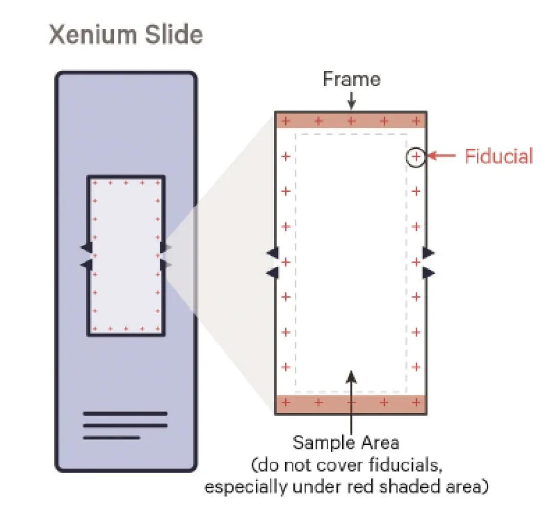
10X Genomics Xenium
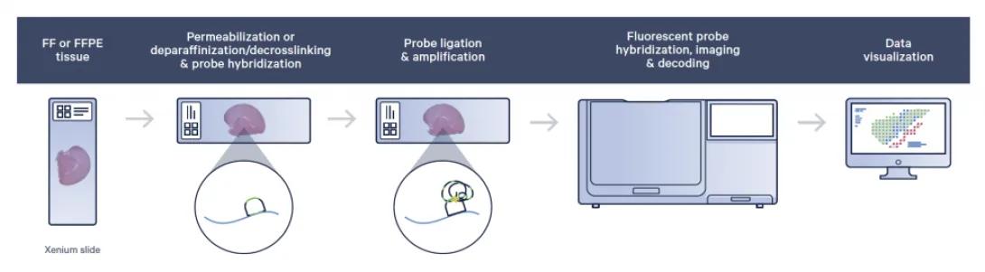
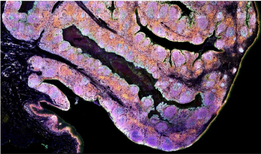
FF or FFPE tissue is sectioned onto a Xenium slide (each slide has an imageable area of 12mm x 24mm). Sections are treated to access the RNA for labeling with circularizable DNA probes. Probe ligation generates a circular DNA probe which is enzymatically amplified. Slides are placed in the Xenium Analyzer where the sample undergoes successive rounds of fluorescent probe hybridization, imaging, and removal; creating bright, easy to image signal with a high signal-to-noise ratio.
An optical signature specific to each gene is generated, enabling target gene identification. Finally, a spatial map of the transcripts is built across the entire tissue section. Data is processed with the 10x Xenium analysis software and can be visualized with the Xenium Browser software.
Sample Requirements
Submission Options
- Tissue processed following the recommended protocols can be submitted directly to the AGC for sectioning and Xenium processing.
- Investigators section tissue onto the Xenium slide. Once tissue is placed on the slide, the slide is stable for up to four weeks, so it can be easily transported to the AGC.
Slide Configuration
Panels
Available now:
- Human Brain
- Human Breast
- Human Colon
- Human Immuno-Oncology
- Human Lung
- Human Multi-Tissue and Cancer
- Human Skin
- 5K Human Pan Tissue & Pathways Panel
- Mouse Brain
- Mouse Tissue Atlas
- 5K Mouse Pan Tissue & Pathways Panel
- Fully Custom – up to 480 genes
Note: Up to 100 custom targets can be added to pre-designed panels
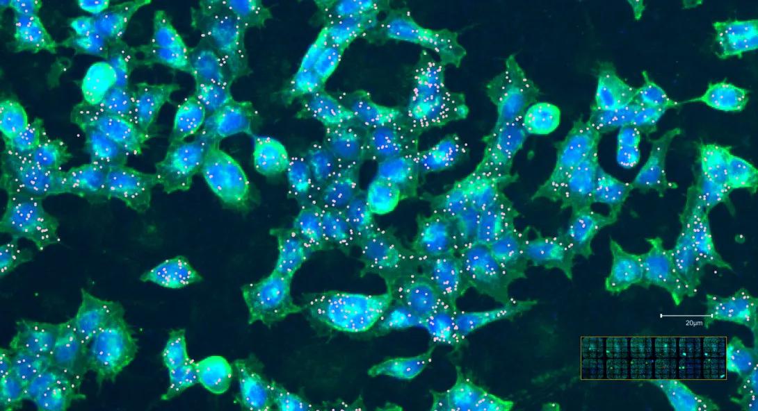
Cell Segmentation
The Xenium with Cell Segmentation Staining method uses a specialized stain mix to precisely identify cell boundaries. These labels facilitate automatic cell segmentation, providing clear boundary definition when membrane signals are pronounced and alternative strategies for boundary identification when membrane staining is unclear. The Xenium Multi-Tissue Stain Mix includes four labeling agents targeting cell membranes, cytoplasm, ribosomal RNA, and nuclei with DAPI.
Cell Segmentation Antibodies
- Membrane markers: E-cadherin (Cdh1), ATPase1a1, CD45
- Cytoplasmic RNA: 18s
- Cytoplasmic proteins: vimentin, aSMA
Cell Segmentation is automatically included with 5K Pan Tissue & Pathways Panels and can be added to any other Xenium pre-designed or custom panel by request.
BRUKER SPATIAL BIOLOGY CELLSCAPE
CellScape enables high-definition protein mapping and quantitative single-cell phenotyping, providing detailed insight into the cellular and molecular architecture of tissues. By integrating advanced spatial resolution with quantitative proteomics, the system supports comprehensive exploration of tumor microenvironments, immune landscapes, and biomarker expression.
Element Biosciences Aviti24 CytoProfiling
The Teton CytoProfiling workflow simultaneously measures transcripts, proteins, and morphology in up to a million cells at subcellular resolution for deep single-cell multiomic profiling.
Plexing
Frequently Asked Questions
Can I practice growing cultures on a Teton Slide?
Yes! You can use the Teton Optimization Kit to optimize cell culture conditions. This kit includes two 12-well flow cell assembly kits and various optimization reagents. Email [email protected] to purchase the optimization kit.
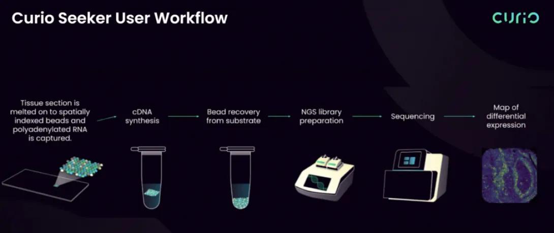
Curio Bioscience Curio Seeker
Curio Seeker tiles are 3 x 3 mm or 10 x 10 mm and contain ~10 µm barcoded beads. The spatial barcode assigned to the bead is incorporated during cDNA synthesis and enables gene expression data to be mapped back to its location within the tissue. Data is processed with the Curio Bioscience analysis software.
Sample Requirements
Fresh/Frozen tissue
- Freshly obtained tissue should be snap frozen to prevent RNA degradation
- The recommended freezing method uses an isopentane and liquid nitrogen bath
- Frozen tissue should be embedded in Optimal Cutting Temperature (OCT), which can be done simultaneously with the freezing process
- RNA quality of the tissue block should be assessed prior to sectioning. The RNA Integrity Number (RIN) should be ≥ 7
Curio Seeker Workflow
Curio Seeker is a set of slides, reagents, and software tools that enable a person to do whole transcriptome analysis on a tissue section. Thus, there are a variety of ways you can opt to use the platform and work with the Advanced Genomics Core.
Tissue Submission – Tissue processed following the recommended protocols can be submitted directly to the AGC for sectioning, staining/imaging, library prep, sequencing, and data processing. The cryomold used for embedding should be of appropriate size to fit the tissue sample. Preferred size for cryoblock submission is 10 mm.
cDNA Submission – Investigators section process onto the tile, perform the Curio assay, and submit the recovered cDNA to AGC. AGC staff will perform library prep, sequencing, and Curio Bioscience pipeline data processing.
Self Service – The Curio Seeker platform does not require specialized instrumentation. Reagents can be purchased directly from Curio Bioscience, and users can prep samples in their own lab space. These samples are submitted to the AGC as user-made libraries ready for Next Generation Sequencing.
Curio Trekker
Curio Trekker's Slide-Tags Methodology
The Curio Trekker Platform employs the innovative Slide-Tags approach, capturing the rich spatial transcriptomics data from intact tissue sections. By tagging single nuclei with spatial barcode oligonucleotides tied to DNA-barcoded beads, Curio Trekker offers precise spatial resolution for single-nucleus profiling.
Key Features:
- Spatial Barcode Technology: Assigns spatial coordinates to individual nuclei via DNA barcode tags, seamlessly integrating with 10x Genomics 3' Gene Expression and Multiome assays.
- Flexible Assay Integration: Versatility in assay selection facilitates comprehensive single-nucleus profiling, encompassing gene expression and chromatin accessibility, while preserving spatial context.
- Species Agnosticism: Curio Trekker's capabilities are not limited by species, providing a universal tool for diverse research applications.
- Tissue Compatibility: Optimized for fresh frozen, OCT-embedded tissues, ensuring the structural integrity and quality of samples.
- Holistic Imaging: Alignment with histological imaging on serial tissue sections offers a complete view of tissue morphology alongside molecular data.
Curio Trekker is the only platform available at the AGC that allows simultaneous capture of both gene expression and chromatin accessibility data from the same sample, maintaining the spatial information critical for comprehensive biological insights.
Questions?
Contact Us
University of Michigan
2800 Plymouth Rd.
Ann Arbor, MI 48109-2800