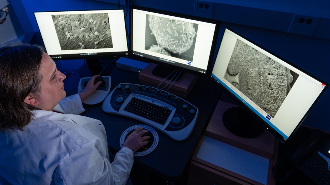
Exceptional sample prep services and access to the latest in electron microscopy
The Microscopy Core houses a mid-range transmission electron microscope (TEM) and scanning electron microscope (SEM) that are suitable for a large majority of biological EM imaging applications. For more specialized techniques, we have partnered with the Engineering School’s Michigan Center for Materials Characterization (MC2) to use their instruments. Core staff will still prepare samples and can collaborate with the (MC)2 staff to assist with imaging.
Are you a New User? Click here for more information on how to get started.
The core offers sample processing services for a wide range of electron microscopy applications. We are also happy to consider method development projects on a collaborative basis. Please contact us to discuss which methods are best suited for your project.
Biological samples like cells and tissues must be properly fixed, embedded in a resin for sectioning, and contrasted with heavy metals prior to TEM imaging. Beginning with chemically fixed specimens, this sample processing service includes washing, secondary fixation, en bloc staining, dehydration, epoxy resin infiltration, embedding and curing. The resultant sample-containing resin blocks can be further sectioned with ultramicrotome for EM imaging (sectioning is provided as a separate service).
Like the fluorescent probes used for protein localization in light microscopy, antibody-conjugated colloidal gold particles are used to label the antigens in electron microscopy with higher resolution. The immunolabeling procedure is conducted on thin sections from samples embedded in LR White resin. The core processes chemically fixed samples into thin sections on Nickel grids for researchers to perform their customized immunolabelling (sectioning is provided as a separate service).
Although chemicals like glutaraldehyde and paraformaldehyde have been used for decades to fix biological samples, this process is associated with artefacts such as structural swelling and distortion. Alternatively, high pressure freezing (HPF) can freeze samples in msecs and preserve them in a “life-like state”. The “frozen” water in samples is then gradually substituted by organic solutions containing fixatives and staining agents. After freeze substitution (FS), samples are gradually warmed up to room temperature and further processed for resin embedding and sectioning as the conventional sample preparation (sectioning is provided as a separate service). Results from HPF-FS show superior preservation of fine structures compared to conventional sample processing.
Similar to the HPF-FS method for ultrastructure study, biological samples are cryo-fixed by HPF, and gradually substituted in organic solutions. Notably, this FS cocktail is designed for immuno-EM to better preserve the antigens. Samples are then infiltrated, embedded, and cured with Lowicryl resin at cryogenic temperatures to further improve the preservation of antigens. The cured blocks are gradually warmed up to room temperature for sectioning (sectioning is provided as a separate service).
Negative-stain TEM is a simple and rapid method to study thin specimens such as viruses, bacteria, exosomes, isolated organelles, and macromolecules. In this method, electron-dense chemicals such as uranyl acetate and phosphotungstic acid are used to stain specimen-adsorbed grids, contrasting the unstained specimen with a dark background.
Prior to SEM imaging, biological samples must be fully dehydrated and applied with a thin conductive coating. To best preserve the surface structures, a state-of-the-art method, critical point drying, is used to dehydrate biological samples. In this method, samples are immersed in liquid CO2 followed by CO2 evaporation at its critical point, where physical characteristics of liquid and gaseous are not distinguishable. Therefore, it avoids damages induced by surface tension when changing from liquid to gaseous state. Next, the dehydrated sample is coated with either carbon or metal for conductivity.
Volume EM allows researchers to visualize a sample slice by slice and create a 3D reconstruction with high resolution. Common ways include focused ion beam -SEM (FIB-SEM), serial block face -SEM (SBF-SEM), and array tomography. The core provides services to process the fixed biological samples to resin blocks for volume EM. The overall procedure is similar to the conventional TEM sample processing but using different protocols for contrasting and embedding. Before the volume EM, the resin blocks can be further trimmed to expose the sample cross-section or confined to a specific area.
Correlative light and electron microscopy (CLEM) is a technique that combines the information acquired from light (LM) and electron microscopy (EM) on the same area of interest from a sample. LM enables multi-color labelling for live and fixed samples in aqueous environment while its spatial resolution is limited. It can be complemented by the high-resolution ultrastructural visualization using EM when studying the same location. This superposition of LM and EM provides a more comprehensive and straightforward information for a biological process.
Sectioning of sample-containing resin blocks is indispensable so that samples are thin enough for TEM visualization. Regular TEM resin sectioning is performed at room temperature using diamond knives to generate sections with thickness of 50-90 nm. Thick sections (>150 nm) can also be performed to accommodate specific project requests. In addition, sectioning at cryogenic temperatures is available. For example, sucrose and gelatin infiltrated samples can be sectioned at ~-110˚C for Tokuyasu method.
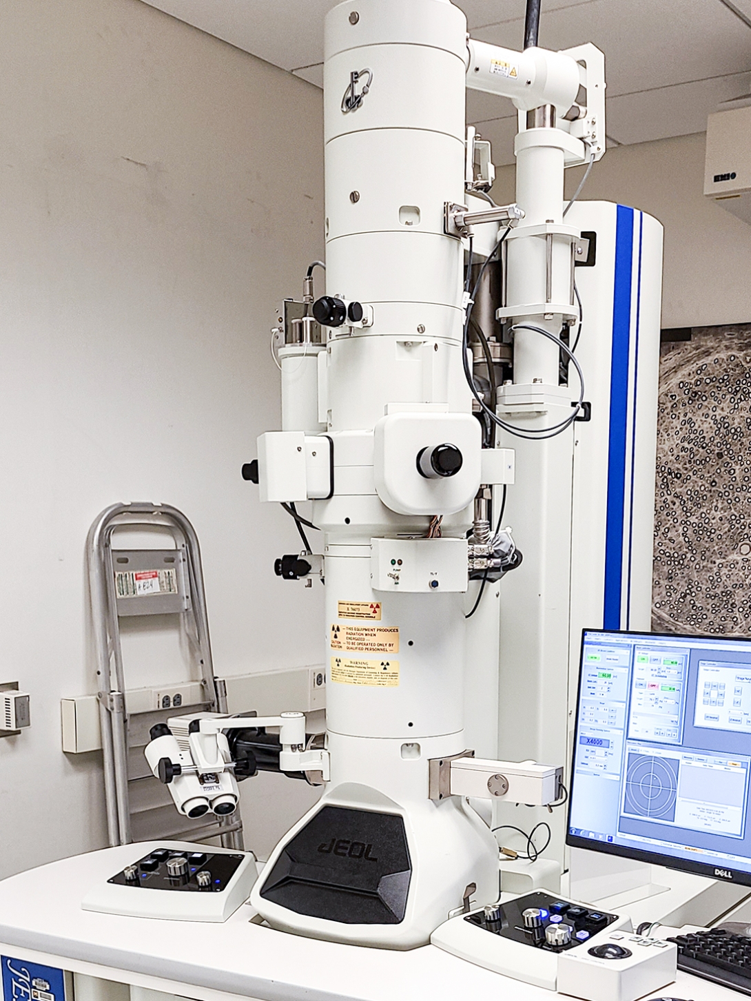
Capabilities: Imaging of thin samples (<200 nm) using transmission electrons
Specifications:
- LaB6 filament
- Resolution: 0.38 nm
- Accelerating voltages: 40 kV to 120 kV
- Magnification: 10x to 1,200,000x
- Equipped with 2k CMOS camera
- Equipped with quick release room temperature retainer EM-11610 QR1 of ±20° tilt
Operations: Users are encouraged to be trained and operate the TEM independently.
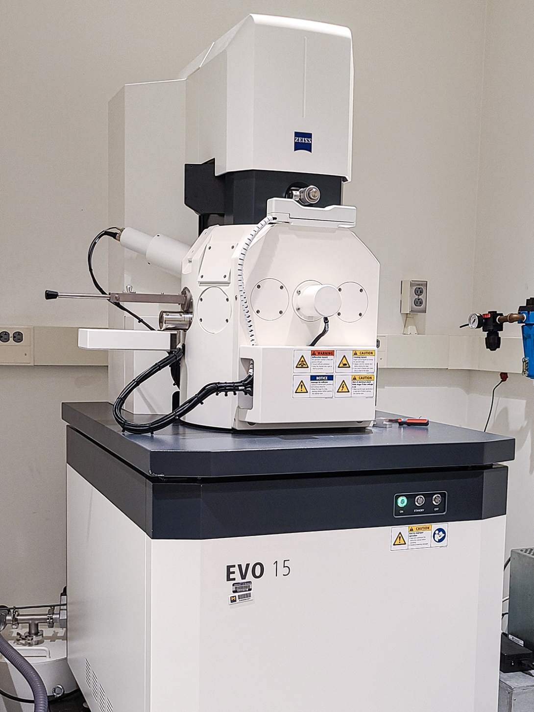
Capabilities: Imaging the surface of dehydrated samples
Specifications:
- LaB6 filament
- Accelerating voltages: 0.2 kV to 30 kV
- Magnification: 5x to 1,000,000x
- Resolution: 2 nm at 30 kV, 8 nm at 3 kV, and 15 nm at 1 kV
- Maximum sample height: 145 mm
- Stage rotation: 360°
- Stage tilt: -10° to +90°
- Equipped with secondary electron detector (SE)
- Equipped with backscattered detector (BSD)
- Equipped with cascade current detector (C2D)
- Equipped with scanning transmission electron microscopy detector (STEM)
Operations: Users are encouraged to be trained and operate the SEM independently.
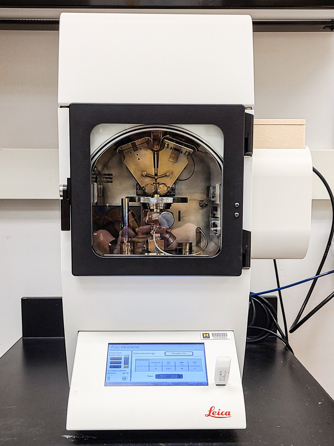
Capabilities:
- Glow discharging substrates and grids
- Sputtering coating surfaces with gold or gold-palladium
- Carbon coating substrates, grids, and sample surfaces
Specifications:
- Oil-free high vacuum system equipped with turbomolecular pump
- Equipped with quartz crystal to continuously monitor coating thickness
- Rotary stage for even distribution of coating materials
- Adjustable stage height and tilt during processing
- Stage that holds two 76 mm x 26 m glass slides or 24 SEM pin stubs
Operations: This instrument is typically operated by BRCF staff. Training may be available upon request.
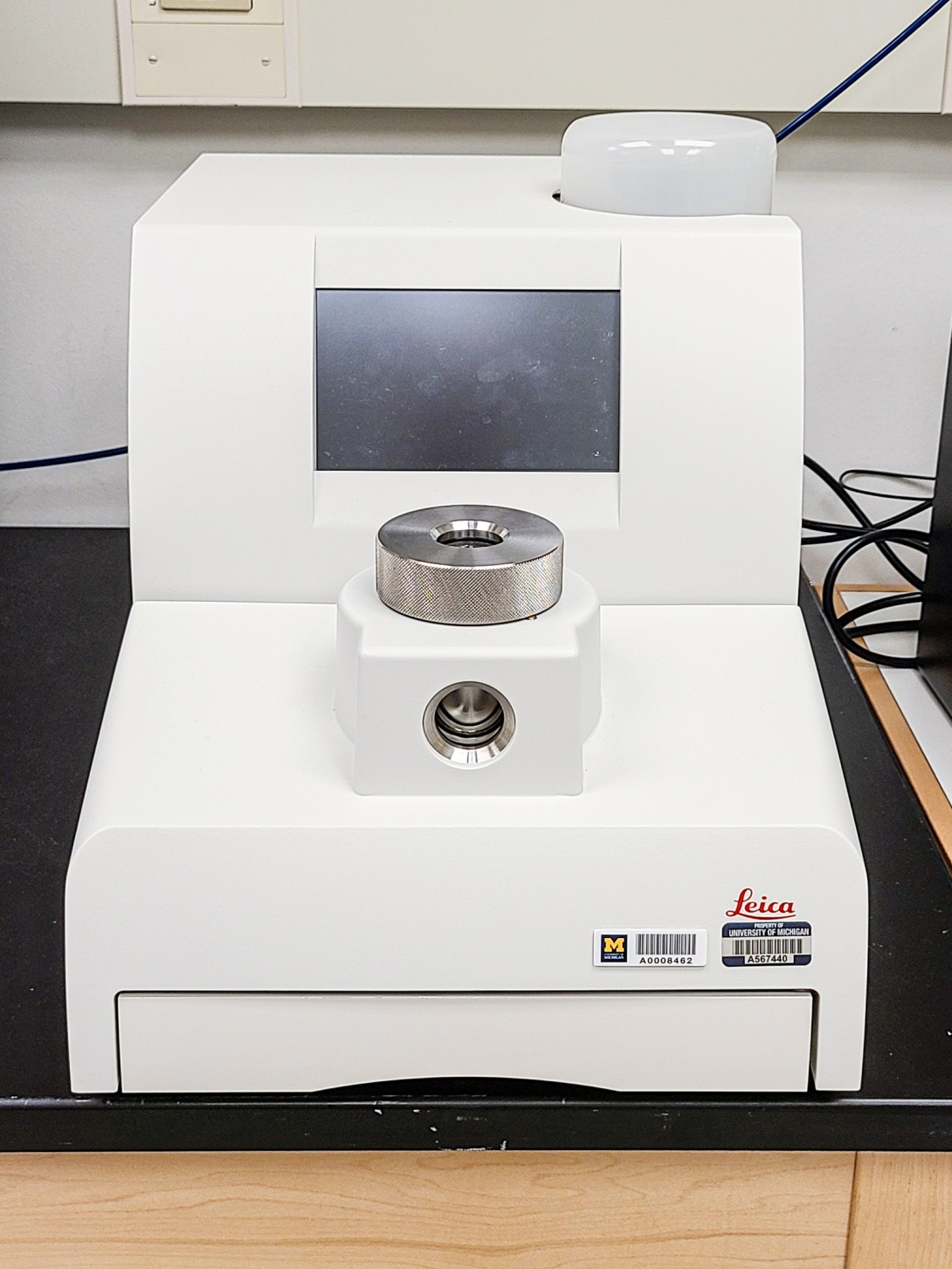
Capabilities: Dehydration of water-containing samples with minimal damages and drying artifacts
Specifications:
- Samples compatible with ethanol or acetone
- Large 60 mm x 62 mm sample chamber and various sample containers
- Maximum operating pressure: 79 bar (CO2 critical point is at 31°C and 73.8 bar)
- Adjustable heating range: 33°C to 43°C
- Adjustable Heating rate: 1°C/min as slow, 2°C/min as medium, 3°C/min as fast
- Adjustable cooling range: 5°C to 25°C
- Safety cutoff for pressure and temperature
Operations: This instrument is typically operated by BRCF staff. Training may be available upon request.
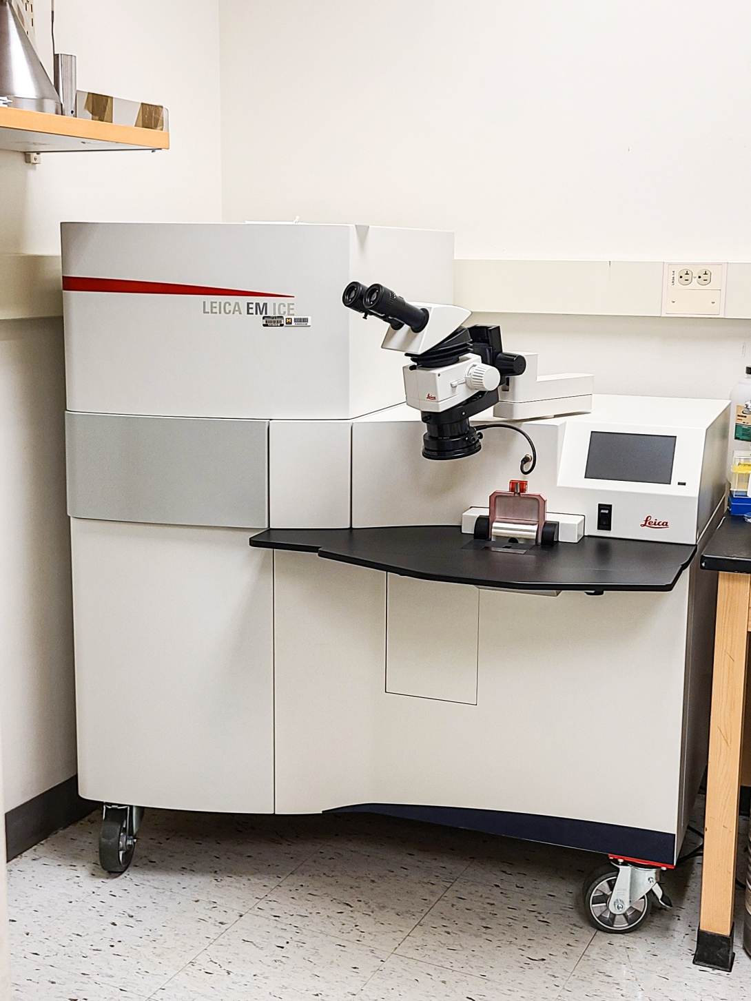
Capabilities:
- Freezing biological samples in milliseconds to preserve delicate structures and transient processes
- Cryo-fixing samples that cannot be chemically fixed, such as liposomes, hydrogels, and micelles
Specifications:
- Cooling rate: 12000-25000 K/s
- Working pressure: 2100 -2300 bar
- Rise time 4-7 ms
- P/T shift: 0-3 ms
- Equipped with 3 mm flat specimen cartridge system
- Equipped with 3 mm sapphire coverslip cartridge system
Operations: This instrument is typically operated by BRCF staff. Training may be available upon request.
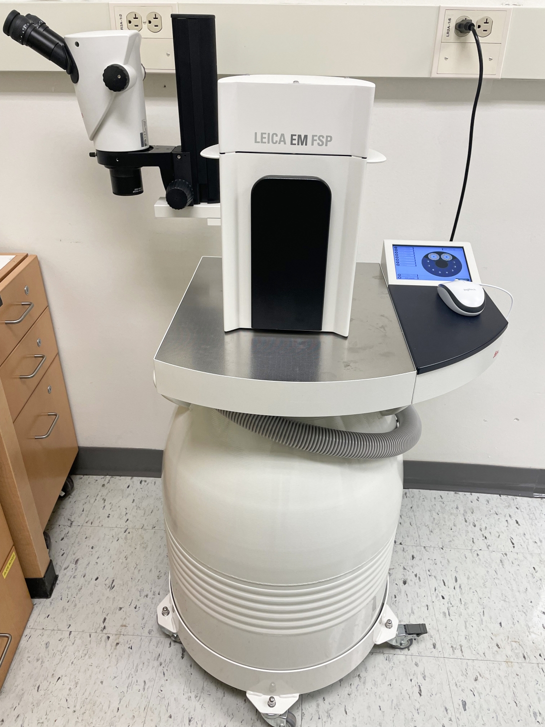
Capabilities:
- Substitution of water in specimens at frozen temperatures
- Resin embedding and UV polymerization at cryogenic temperatures
- Other applications with progressive lowering of temperature
Specifications:
- LN2 dewar: 35 L for 5-day consumption
- Temperature: -140°C to 70°C
- Deep Freeze mode < -140°C
- Setup of 1-99 programmable steps
- TF function that excludes water and oxygen from sample chamber
- Equipped with Leica EM FSP for automated liquid exchange, mix, agitation, and UV polymerization
Operations: This instrument is typically operated by BRCF staff. Training may be available upon request.
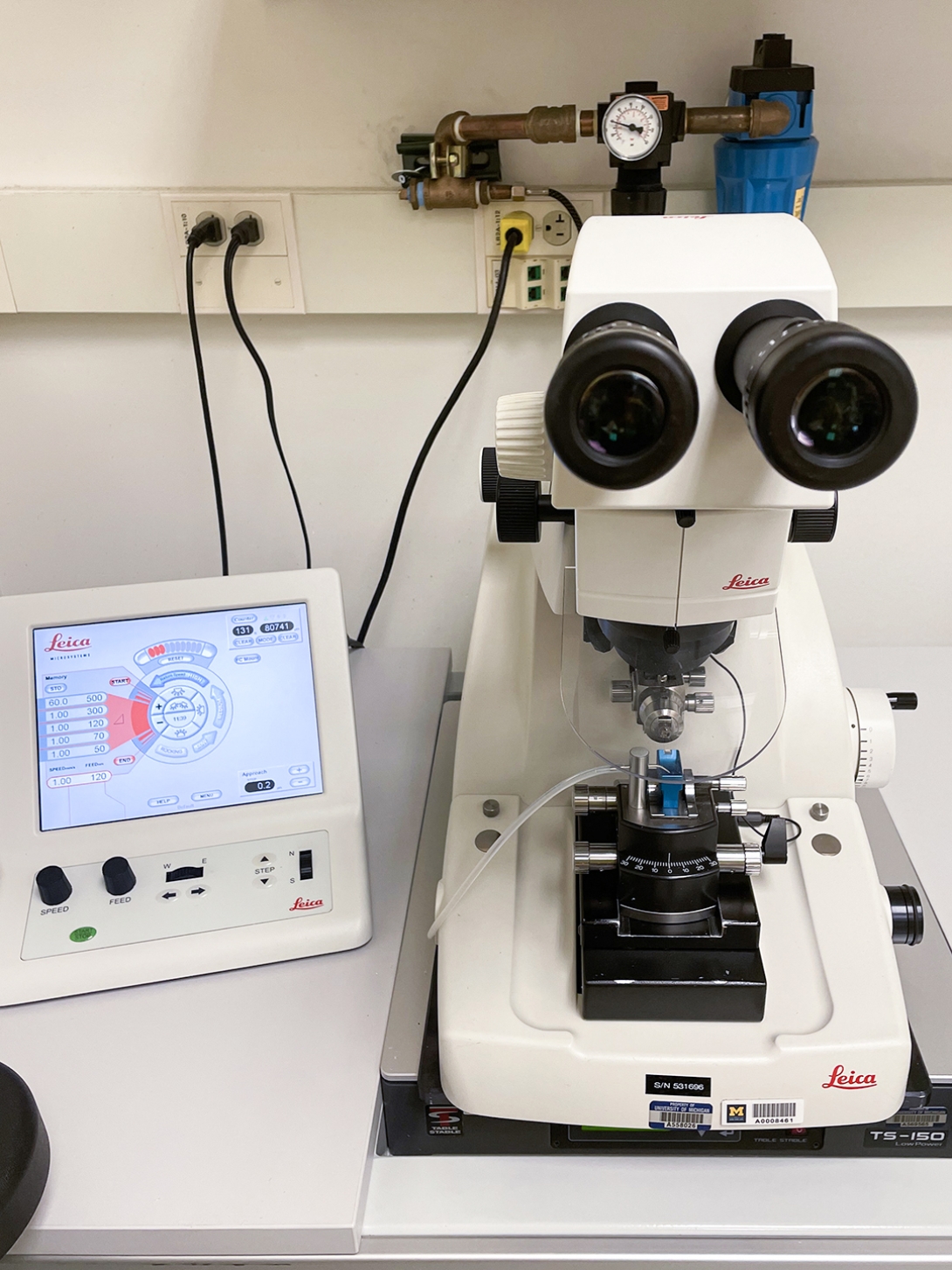
Capabilities: Sectioning soft samples into thin and semithin sections for microscopy
Specifications:
- Specimen advance: 200 µm
- Knife holder: for one 6-12 mm knife
- Clearance angle: -2° to 15°
- Cutting window: 0.2 to 14 mm
- Cutting speed: 0.05 to 100 mm/s
- Equipped with Ultra 45° Diatome for 30-200 nm sections
Operations: This instrument is typically operated by BRCF staff. Training may be available upon request.
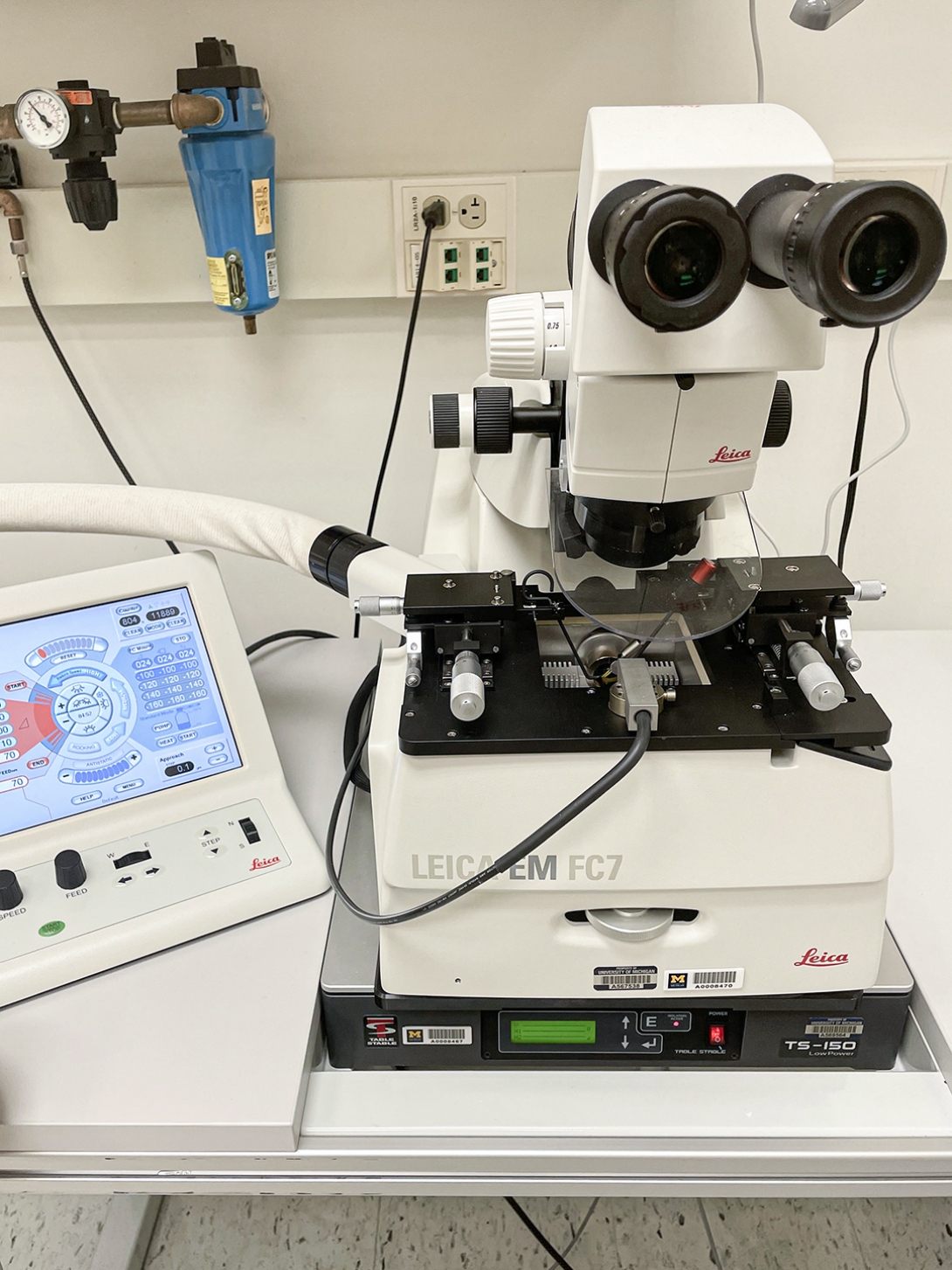
Capabilities: Sectioning soft samples at cryogenic temperatures, e.g., frozen biological samples and rubbers
Specifications:
- Sectioning temperature: -15° C to -185°C
- Temperature difference setting for knife/sample: -40°C for knife and -170°C for sample
- Knife holder: for two 6-10 mm knives
- Equipped with Leica EM CRION ionizer for electrostatic charge and discharge functions
- Equipped with micromanipulator for section collection
Operations: Serial sectioning of soft samples for array tomography 3D reconstruction from EM section images
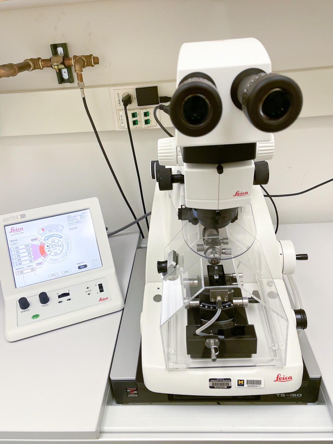
Capabilities: Serial sectioning of soft samples for array tomography 3D reconstruction from EM section images
Specifications:
- Specimen advance: 200 µm
- Knife holder: for one 6-12 mm knife
- Clearance angle: -2° to 15°
- Cutting window: 0.2 to 14 mm
- Cutting speed: 0.05 to 100 mm/s
- Equipped with AT4 knife of 4 mm diamond blade
Operations: This instrument is typically operated by BRCF staff. Training maybe available upon request.
All New Users of Electron Microscopy must complete the New Project Request Form and enroll in MiCores. Upon completion, the Microscopy team will be in contact shortly.
How to Enroll in MiCores
- Log in to MiCores using your uniqname and Level-1 password.
- Add your affiliation with a PI or Lab Manager. Your PI or Lab Manager must provide you access to one or more shortcodes.
- For questions on these steps, please contact the HITS Help Desk, or visit the MiCores Learning Site.
Building Access Requests
To access the BSRB core after hours, or the NCRC core at any time:
- Complete Bloodborne Pathogens (EHS_BLS101W or BLS100W) and General Laboratory Safety (BLS025W) and email the online learning certificates to brcf-coreaccess@umich.edu.
- In the e-mail, include the following information:
- Affiliation (Medical School, College of Engineering, LS&A, etc.)
- Employee type (faculty, staff, or student)
- Access requested (NCRC or BSRB core? Is building door access also needed?)
Please note: Access requests take approximately 72 hours to complete.