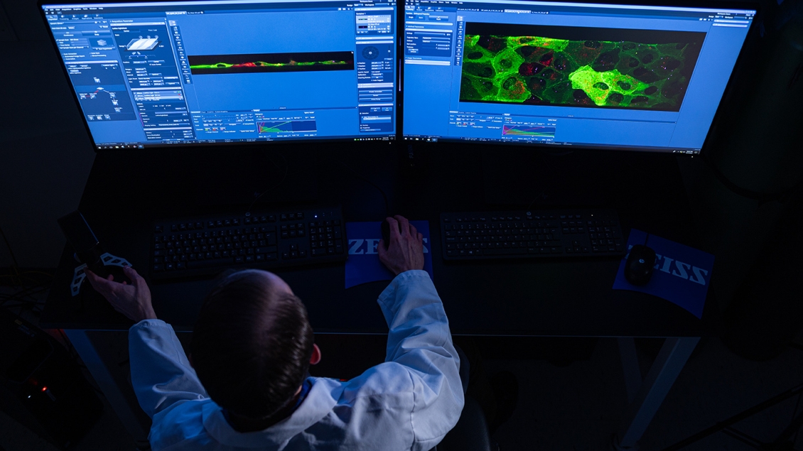
Advanced light microscopes and imaging systems available for your research
The BRCF Microscopy Core houses a wide range of light microscopes and imaging systems in the following three locations: BSRB A830, MSII 5631, and NCRC B20 57S. These instruments support a variety of imaging modalities, as described below. Please contact us during the planning stage of your project to help you select the most appropriate methodology for your research needs.
Are you a new user? Learn how to get started.
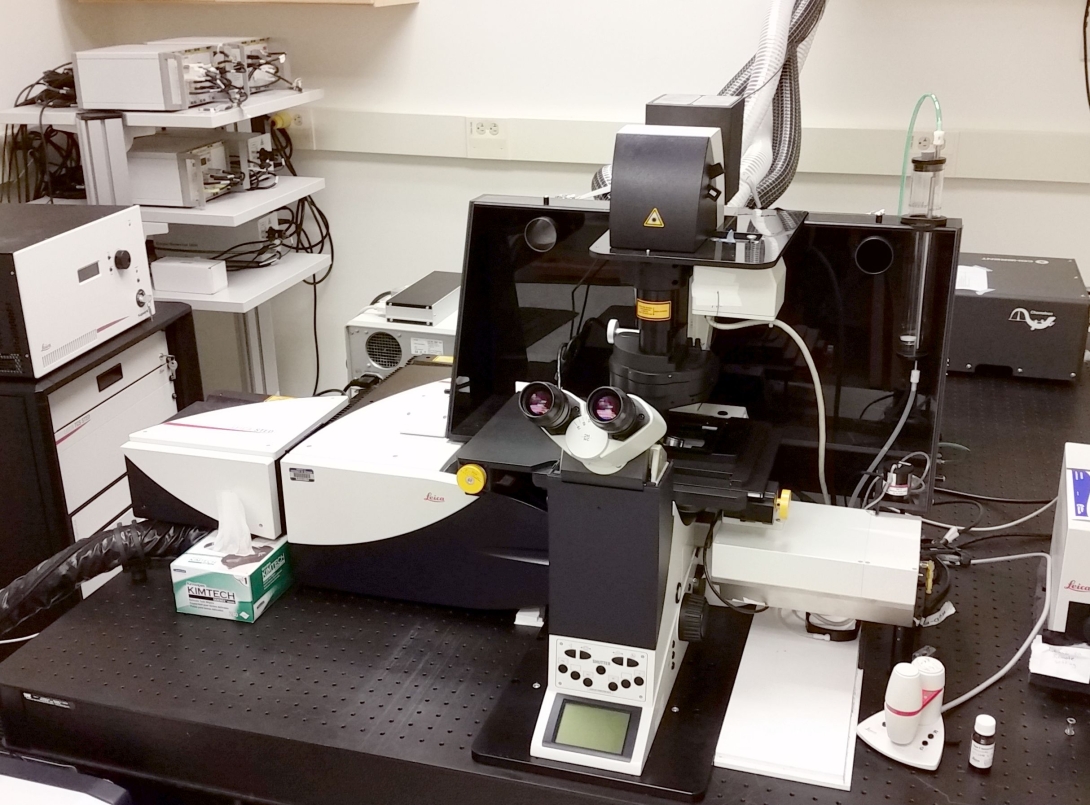
Laser Scanning Confocal, Multi Photon, Super Resolution
- A high sensitivity, inverted, point-scanning confocal and multiphoton system, suitable for fixed and live samples.
- Equipped with a resonant scanner for fast image acquisition.
- Capable of Fluorescence Lifetime Imaging (FLIM).
- Single 594 nm depletion line for Stimulated Emission Depletion Microscopy (STED) in XY only.
- Lasers available for imaging:
- 405nm diode laser.
- Argon laser with 5 excitation lines (458nm, 476nm, 488nm, 496nm, 514nm).
- White light laser tunable from 470nm-670 nm.
- Ti: Sapphire pulsed IR laser (100 fsec, 80 MHz). Tunable from 670nm-1080nm.
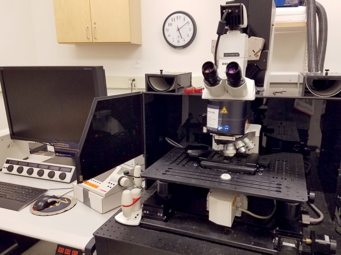
Laser Scanning Confocal, Multi Photon
- A high sensitivity, upright, point-scanning confocal and multiphoton system, equipped with a large stage and dipping lenses.
- Equipped with a resonant scanner for fast image acquisition.
- Lasers available for imaging:
- 405nm Diode laser.
- Argon Laser with 5 excitation lines (458nm, 476nm, 488nm, 496nm, 514nm)
- 561nm diode laser
- 633nm HeNe laser
- Ti: Sapphire pulsed IR laser (100 fsec, 80 MHz). Tunable from 670nm-1080nm
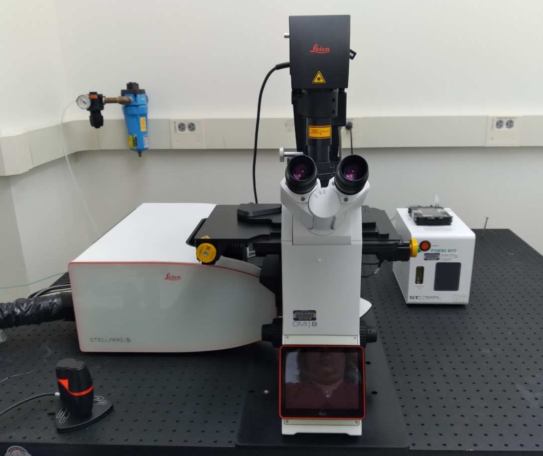
Laser Scanning Confocal
- A high sensitivity, inverted, point-scanning confocal system, suitable for fixed and live samples.
- Equipped with a resonant scanner for fast image acquisition.
- Uses Navigator software for easy stitching, tiling, and overview imaging.
- TauSensing tools, including TauContrast, TauGating and TauSeparation are available. These tools use the differences in fluorescence lifetimes to separate closely overlapping fluorophores and suppress nonspecific background fluorescence.
- 405nm diode laser.
- 445nm diode laser.
- White light laser tunable from 470nm-680 nm.
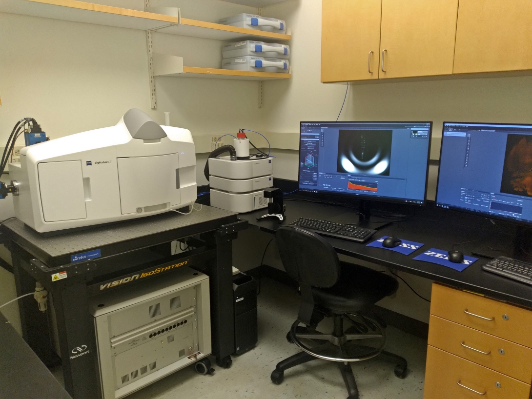
Light Sheet
- Gaussian light sheet instrument, ideal for imaging larger format samples including organoids, zebrafish, embryos, and cleared murine organs.
- Dry and immersion objectives corrected for RI = 1.33 – 1.58; 2X, 5X and 20X objectives.
- 6 available laser lines: 405nm, 445nm, 488nm, 515nm, 561nm and 647nm.
- Incubation for live timelapse imaging.
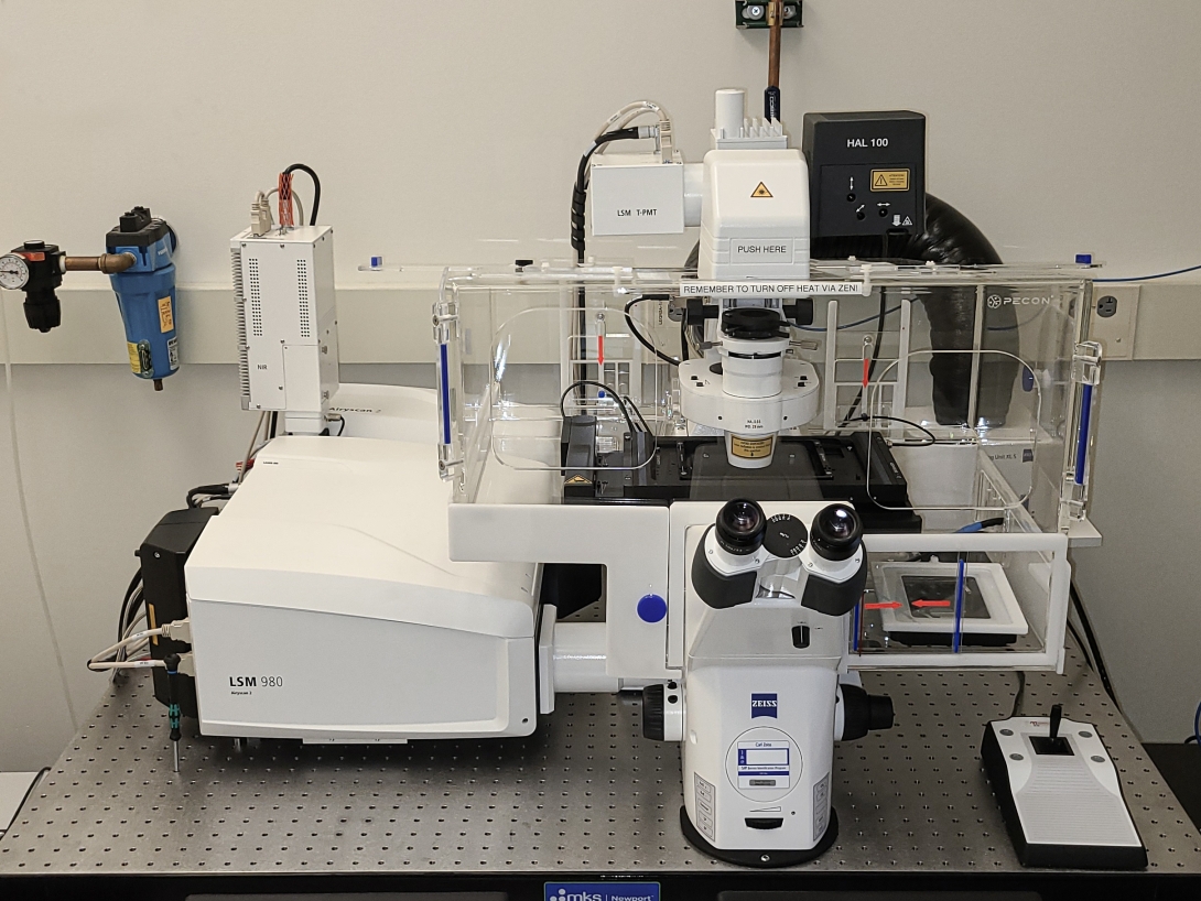
Laser Scanning Confocal, Super Resolution
- A high sensitivity inverted point scanning confocal microscope equipped with an Airyscan detector capable of super-resolution imaging of fixed and live samples.
- Equipped with a spectral detector for confocal imaging of fluorophores with highly overlapping spectra.
- Incubation for timelapse imaging.
- Available laser lines:
- Confocal: 405nm, 488nm, 561nm, 639nm, and 730nm
- Airyscan: 405nm, 488nm, 561nm, and 639nm
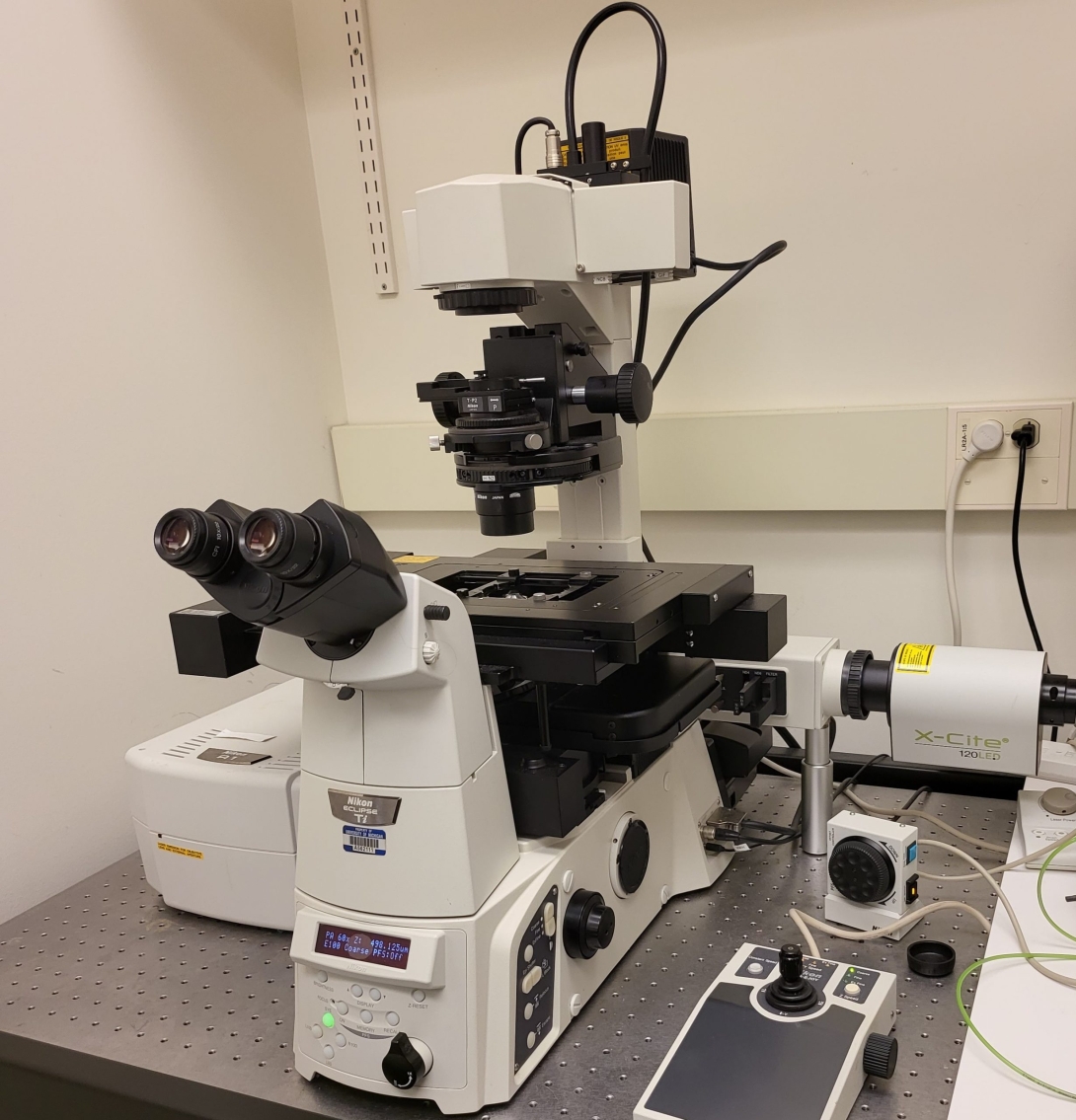
Light Scanning Confocal
- An inverted point scanning confocal with two standard PMT detectors and two GaAsP detectors.
- Ideal for slides and other fixed sample formats. Basic live cell imaging is possible.
- 4 available laser lines: 405nm, 488nm, 561nm and 647nm
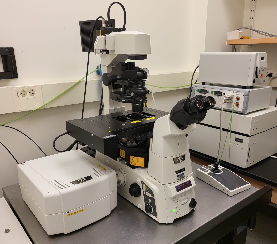
Light Scanning Confocal
- An inverted point scanning confocal with standard PMT detectors.
- Ideal for slides and other fixed sample formats. Basic live cell imaging is possible.
- 4 available laser lines: 405nm, 488nm, 561nm and 647nm
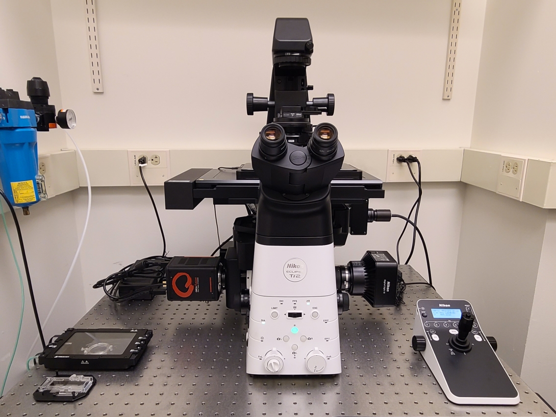
Widefield
- Inverted widefield microscope supporting the following modalities: brightfield, fluorescence, DIC, phase contrast.
- Eight fluorescence channels for blue, cyan, green, yellow, orange, red, far-red and near IR fluorophores.
- Fully motorized to facilitate a variety of experiments including large image stitching.
- NIS Elements software including JOBS, HCA and GA3 modules for advanced acquisition protocols and some analyses in real time.
- Incubation for live timelapse imaging.
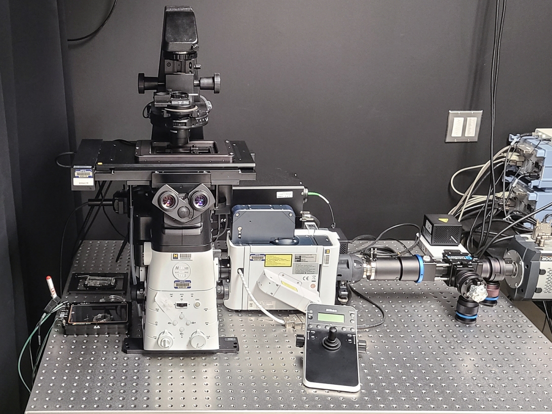
Spinning Disk Confocal, Total Internal Reflection Fluorescence, Widefield
- An inverted spinning disk confocal system, ideal for fast, gentle imaging of live samples such as cells in culture and small organoids and organisms. Speeds of 30 fps possible.
- Digital mirror device for photo-activation or optogenetics using a 395nm or 470nm LED.
- Through objective TIRF illumination available. Monochrome camera for TIRF and widefield imaging.
- 4 available laser lines for confocal (3 for TIRF): (405nm), 488nm, 561nm and 647nm.
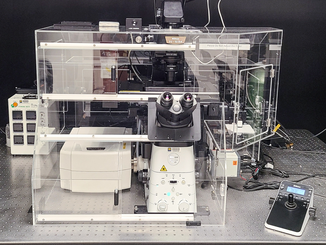
Laser Scanning Confocal, Super Resolution, Widefield
- An inverted microscope equipped with a point-scanning confocal and a 2D / 3D structured illumination (SIM) system.
- Equipped with a resonant scanner for fast confocal image acquisition.
- Timelapse widefield live-cell imaging is also supported.
- 6 available laser lines for confocal (4 for SIM): 405nm, (445nm), 488nm, (514nm), 561nm and 647nm.
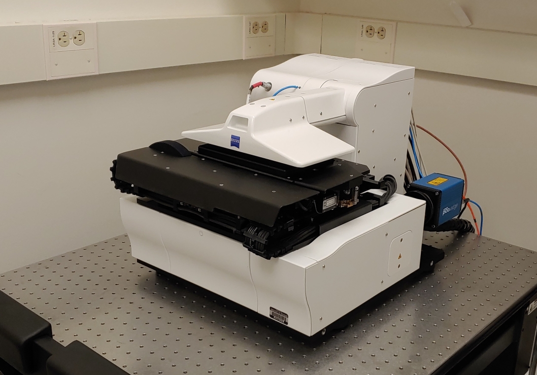
Light Sheet
- Fast and gentle volumetric imaging with minimal photobleaching and phototoxicity.
- Ideal for long-term imaging of dynamic subcellular processes in living cells. Not recommended for samples greater than 50um thick.
- 40X objective; 3 laser lines: 488nm, 561nm, and 633nm
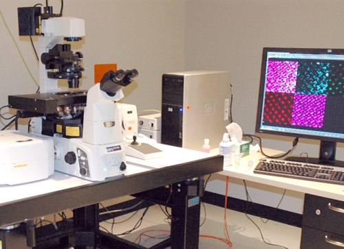
Laser Scanning Confocal
- An inverted point scanning confocal, suitable for live and fixed samples
- Includes a spectral detector for imaging dyes with highly overlapping emission profiles
- 4 available laser lines: 405nm, 488nm, 561nm and 647nm
Spinning Disk Confocal, Widefield
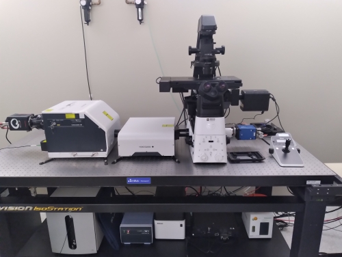
- An inverted spinning disk confocal system, ideal for fast, gentle imaging of live samples
- Wide field of view, enabling fast tiling of live and fixed samples
- SoRa mode available, offering 2X improvement in resolution by pixel reassignment
- 4 available laser lines: 405nm, 488nm, 561nm and 647nm
Tissue Clearing
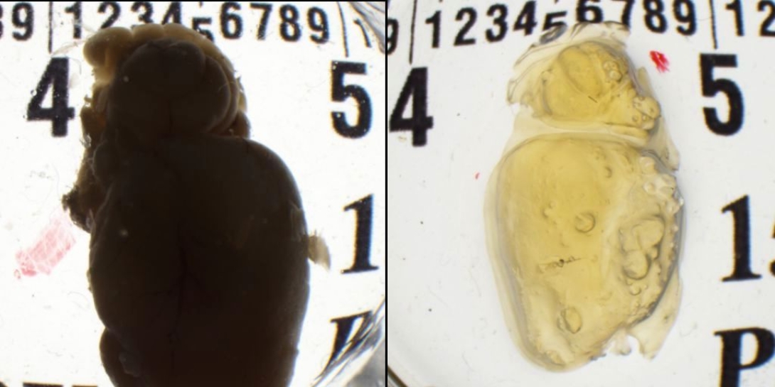
Clearing protocols make tissue highly transparent by removing lipids and refractive index matching, enabling deep imaging through large volumes of tissue. We offer iDisco clearing on a fee-for-service basis and can advise and assist with other organic solvent-based clearing protocols. Additionally, the core houses the LifeCanvas systems for electrophoretic clearing and staining via methods including CLARITY and SHIELD. Please contact us for further information.
Expansion Microscopy
Expansion microscopy cross links the proteins in the sample to a polymer backbone and then swells the polymer mesh to make the sample larger (usually by 4x in each dimension), thus enabling super resolution imaging on a standard fluorescence microscope. We offer the MAP and proExM protocols on a fee-for-service basis. Please contact us with questions.
All New Users of Light Microscopy must complete the New Project Request Form and enroll in MiCores. Upon completion, the Microscopy team will be in contact shortly.
How to Enroll in MiCores
- Log in to MiCores using your uniqname and Level-1 password.
- Add your affiliation with a PI or Lab Manager. Your PI or Lab Manager must provide you access to one or more shortcodes.
- For questions on these steps, please contact the HITS Help Desk, or visit the MiCores Learning Site.
Building Access Requests
To access the BSRB core after hours, or the NCRC core at any time:
- Complete Bloodborne Pathogens (EHS_BLS101W or BLS100W) and General Laboratory Safety (BLS025W) and email the online learning certificates to brcf-coreaccess@umich.edu.
- In the e-mail, include the following information:
- Affiliation (Medical School, College of Engineering, LS&A, etc.)
- Employee type (faculty, staff, or student)
- Access requested (NCRC or BSRB core? Is building door access also needed?)
Please note: Access requests take approximately 72 hours to complete.