Microscopy Core
Offering state-of-the-art microscopy instrumentation and services to support our researchers.
About the Microscopy Core
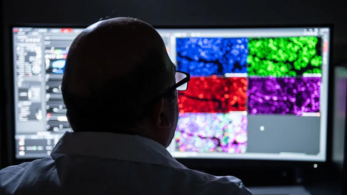
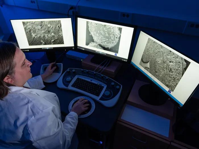
The Microscopy Core offers support and training on a variety of high-end instrumentation and advanced methodologies for both light and electron microscopy.
The core also offers a wide range of sample preparation services for electron microscopy, and training and assistance with image analysis.
Need help?
- Request a project, consultation, or training with Microscopy Core experts.
- Are you a new user or need card reader access? Learn how to get started.
- All other inquiries should be directed via email to the Microscopy Core. Please to not use this e-mail to inquire about microscope training. Fill out the form instead.
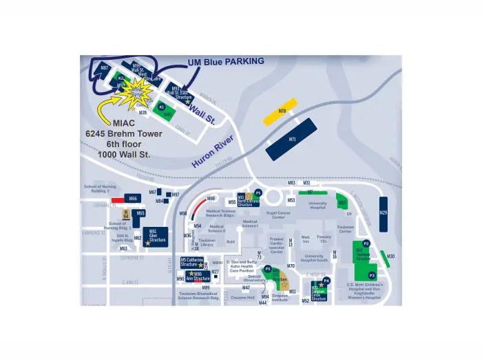
MIAC is Now Part of the BRCF Microscopy Core
We are pleased to announce that the BRCF Microscopy Core is extending our footprint to the Brehm Tower. All users will be able to book microscopy instrumentation and services at Brehm as well as at the three existing Microscopy Core locations (BSRB, MSII and NCRC) through the BRCF Microscopy Core iLabs Site. Current MIAC users will experience the same high level of service and scientific leadership from Dr. Stephen Lentz, who will continue to manage the Microscopy Core site at Brehm.
A new Nikon AXR NSPARC laser scanning confocal and superresolution microscope has been added to the BRCF Microscopy Core site in MSII, and this system has replaced the Nikon A1R + SIM which has been moved and reinstalled at Brehm.
Additional information on our imaging systems can be found on the light microscopy page of our website.
How We Serve Your Research Needs
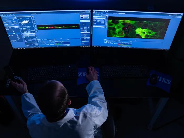
Light Microscopy
The Microscopy Core houses a wide range of light microscopes and imaging systems. These instruments support a variety of imaging modalities.
Electron Microscopy
The Microscopy Core owns a basic transmission electron microscope (TEM) and scanning electron microscope (SEM) that are suitable for a large majority of biological EM imaging applications.
Image Analysis
In addition to Light Microscopy and Electron Microscopy services, the Microscopy Core provides researchers with support across a variety of open-source and commercial image analysis software packages.
Open Source Software:
Commercial Software:
Almost any feature or quality that can be imagined, such as number, size, shape, or intensity, can potentially be measured. Common image analysis requests include: Multi-channel Segmentation, Object Counting, Object Tracking, and 3D Rendering and Visualization.
If you are interested in image analysis, guidance, or education, please contact us.
Our Rates
Rates and fees for routine services are listed below. For complex projects that require extensive labor, please consider including core staff as key personnel on grant submissions. Contact us with any questions you may have.
Rates are subject to change following Office of Financial Analysis review.
| Device | Internal Peak | Internal Off-Peak | Overnight | External Academic | External Industry |
| All confocal, superresolution and lightsheet microscopes | $42/hr | $32/hr | $10/hr | $58/hr | $70/hr |
| Ti2 Widefield Microscope | $24/hr | $18/hr | $6/hr | $35/hr | $40/hr |
| Zeiss EVO 15 SEM & JEOL JEM-1400+ TEM | $42/hr | $32/hr | N/A | $58/hr | $70/hr |
| Leica EM UC7 Ultramicrotome* | $25/hr | $25/hr | N/A | $35/hr | $50/hr |
| Imaris | $8/hr | $8/hr | N/A | $12/hr | $20/hr |
For all instruments except the Yokogawa CellVoyager 8000, peak times are Monday – Friday, 8:00 am – 6:00 pm. For the Yokogawa CV8000, peak times are 8:00 am Monday – 8:00 am Saturday. All other times are Off-Peak. For overnight time lapse experiments over 12 hours only, the time between 12:00 am – 8:00 am can be billed at the overnight rate. Please inquire regarding the cost and scheduling for time lapse experiments lasting more than 24 hours.
*Skilled users can use an ultramicrotome if they bring their own diamond knife. Training in ultramicrotomy is not typically provided, but could be offered to interested users and would be charged at the labor rate.
| Service | Internal | External Academic | External Industry |
| Training Per Hour Charge | $75 | $105 | $120 |
| Conventional EM Sample Preparation (1-4 samples) | $415 | $575 | $700 |
| Advanced EM Sample Preparation (1-4 samples) | $965 | $1,350 | $1,550 |
| Ultramicrotome Sectioning: Mesh Grids (per sample) | $155 | $225 | $250 |
| Ultramicrotome Sectioning: Slotted Grids (per sample) | $205 | $300 | $350 |
| Ultramicrotome Re-sectioning (per block) | $80 | $115 | $140 |
| Agarose embedding surcharge (1-4 samples) | $40 | $55 | $70 |
| Monolayer surcharge (1-4 samples) | $75 | $105 | $135 |
| Post-section staining (1-2 grids/sample) | $15 | $24 | $30 |
| High Pressure Freezing (per session) | $250 | $350 | $450 |
| Negative Staining (per session) | $50 | $70 | $90 |
| Critical Point Dryer (per use) | $100 | $135 | $160 |
| Glow Discharger (per use) | $50 | $70 | $90 |
| Sputter Coater (per use; up to 10 nm) | $50 | $70 | $90 |
| Sputter Coater (per use; each additional 10 nm) | $5 | $7 | $10 |
| Additional EM Labor Per Hour Charge | $75 | $105 | $125 |
| Clearing / Expansion Sample Prep (per sample) | $100 | $150 | $200 |
| Life Canvas Per Use Charge (excludes reagents) | $50 | $75 | $100 |
| Additional LM Labor Per Hour Charge | $75 | $105 | $125 |
| Image Analysis Labor Per Hour Charge | $75 | $105 | $125 |
Training is charged at the instrument rate plus the labor rate. Training typically lasts 2-4 hours but can take more or less time depending on the complexity of the experiment and the needs of the user. Any service not specified in the rate table will be charged at the labor rate.
Learning & Outreach
The Microscopy Core offers resources and workshops to support researchers with image analysis and microscopy. Future events will be added as they are offered. Researchers can also find self-guided resources and presentations from past lectures below.
Facilities & Resources
The BRCF Microscopy Core is a centralized, shared instrumentation facility occupying over 3,000 sq. ft. in the University of Michigan Biomedical Sciences Research Building (BSRB). Smaller satellite sites are in the University of Michigan Medical Sciences II (Med Sci II) building on the medical campus, and at the University of Michigan North Campus Research Complex (NCRC). The core features cutting-edge equipment for both light and electron microscopy and supports facility users’ projects from conception through publication by providing consultation, training, and assistance with data acquisition and analysis. Biological sample preparation and sectioning for electron microscopy are also offered. The core is staffed by six highly trained and experienced imaging scientists with expertise in advanced light and electron microscopy and imaging techniques.
Our Leadership
Gary Luker, MD
Professor, Associate Chair, MM Radiology
Jennifer Peters, PhD
CZI Imaging Scientist
Eric Rentchler, PhD
Light Microscopy Lead
Jing Liang, PhD
Electron Microscopy Lead
Stephen Lentz, PhD
Associate Research Scientist

[email protected]
Binyamin Jacobovitz
[email protected]
Megan Bennett
[email protected]
Questions?
Our Locations
Biomedical Science Research Building
109 Zina Pitcher Drive, Room A830
North Campus Research Complex
2800 Plymouth Rd, Room 53S
Medical Sciences II
1137 Catherine St, Room 5631
Brehm Research Tower
1000 Wall St, Room 6245
About Us
The Microscopy Core is one of the Biomedical Research Core Facilities, and a part of the Medical School Office of Research, where our mission is to foster an environment of innovation and efficiency that serves the Michigan Medicine research community and supports biomedical science from insight to impact.
For more information, please refer to the New User Page or email [email protected]
Upcoming Research Events
Ask Lu Anything: Insights in Single-Cell & Spatial Omics with Dr. Luciano Martelotto
The Funding Forum
FFMI Industry-Ready Webinar Series: IP, Data, and Publications in Industry-Sponsored Research
New England Biolabs Product Showcase
Oncology Drug Discovery & Development (3D) Workshop
The Funding Forum
Researchpalooza
The Funding Forum
The Funding Forum
Featured Research News
New Research Team Collaboration Agreement Template
New Requirement for Dual Appointment VA Faculty Serving as PIs on U-M Sponsored Projects