Islet Core
Advanced Diabetes Research
The Islet Core provides consultative advice and state-of-the-art equipment and services for the isolation and study of pancreatic islet structure and function in support of diabetes-related research.
How We Serve Your Research Needs
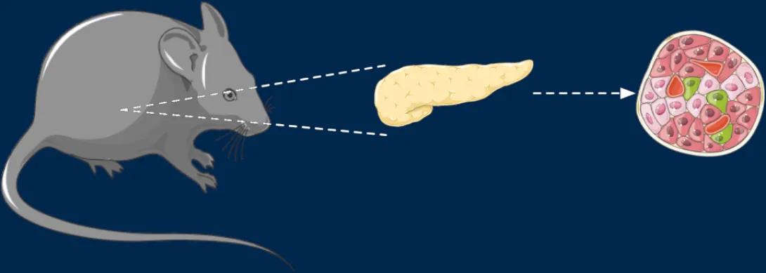
- Pancreas
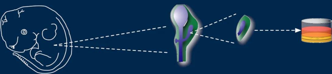
- Embryonic rudiments (E10.5 — birth)
- Islet isolation
Embryonic tissue dissection
Tissue harvesting and islet isolation
- β- α-cell mass
- Proliferation
- Apoptosis
β-cell mass
5 sections spread throughout the pancreas;
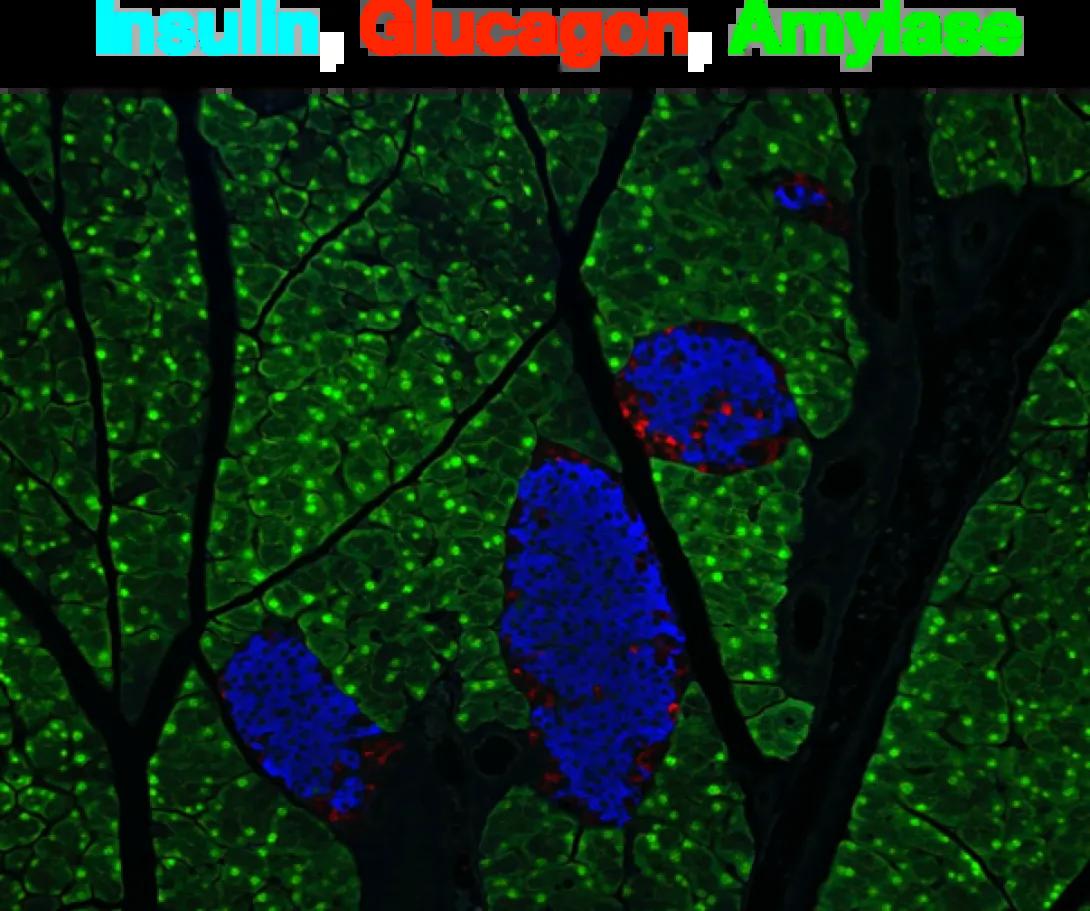
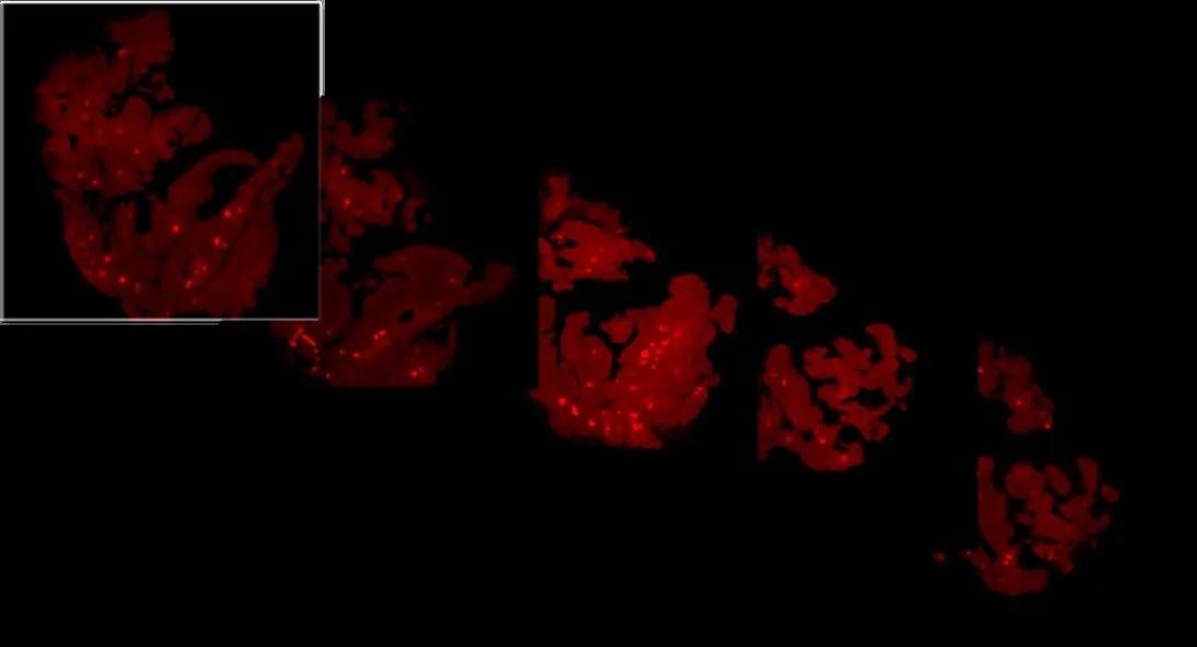
- Fluorescent Immunohistochemistry staining for insulin
- Imaging
- Morphometry

Immunostaining
In addition to insulin and glucagon, a number of antibodies for pancreatic markers are available through the Core.
Software assisted nuclei counting
- Proliferation (Ki67)
- Apoptosis (TUNEL) staining
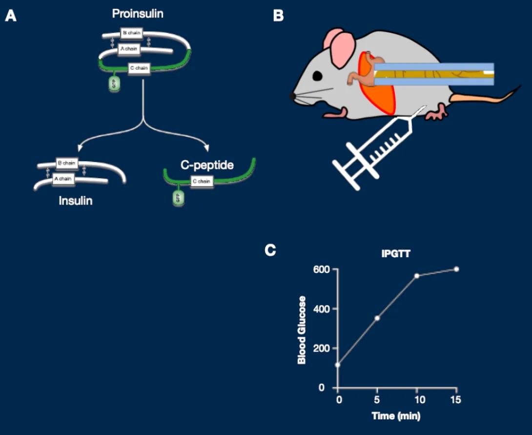
In vivo imaging of fluorescent reporters in anesthetized mice
- Imaging during the course of a glucose tolerance test
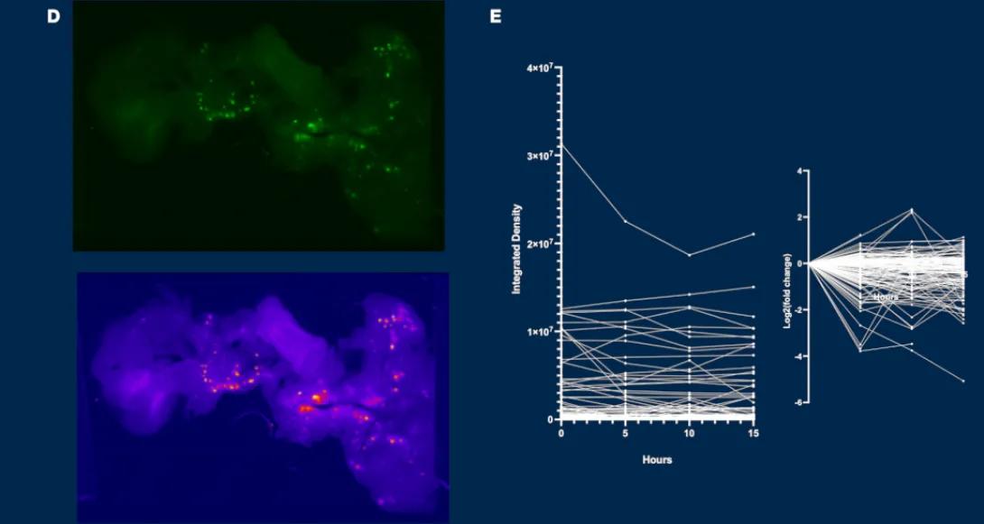
- Image processing for analysis
- Islet tracking over the course of the glucose challenge
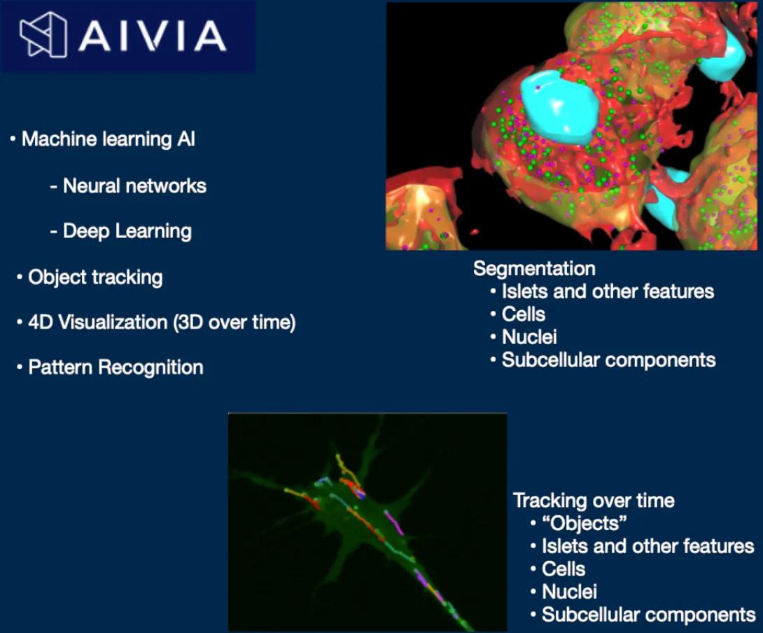
AI-based image analysis
Using Aivia (Microscopy and Imaging Core), we can automate morphometric assessments (β- α-cell mass, proliferation and apoptosis) as well as fluorescent islet tracking and signal quantification after neural network training.
• Static GSIS
• Perifusion
• Respirometry
Static Glucose-stimulated insulin secretion
In culture with transwell inserts

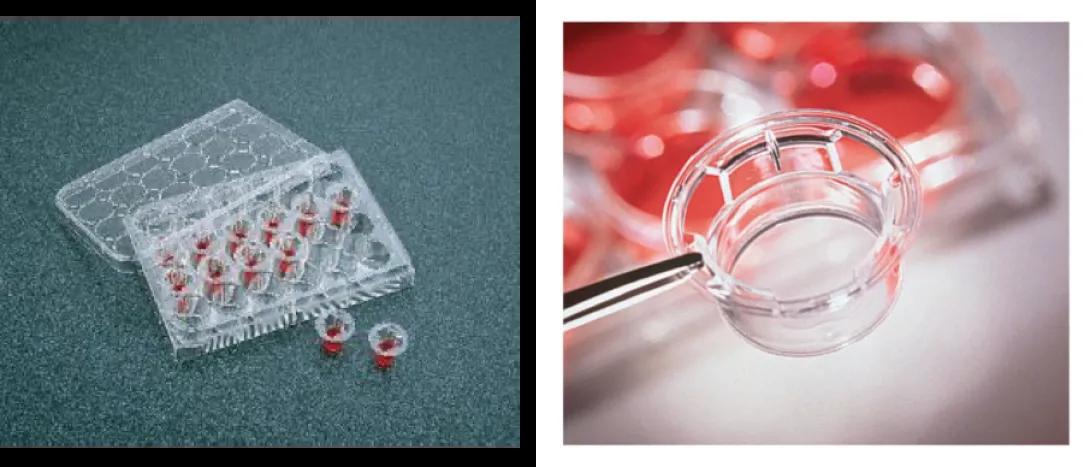
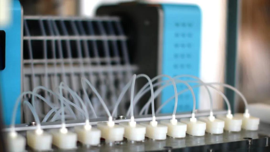
Perifusion
Up to 12 chambers can be used in parallel to measure islet secretions using the custom program of your choice.
The perifusion chambers and reagents are temperature-controlled. The reagents can be gazed with CO2.
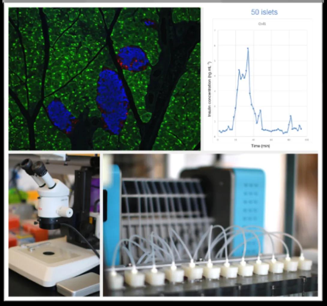
Respirometry (BaroFuse)
Respirometry and perifusion can run concomitantly on up to 6 samples at the same time.
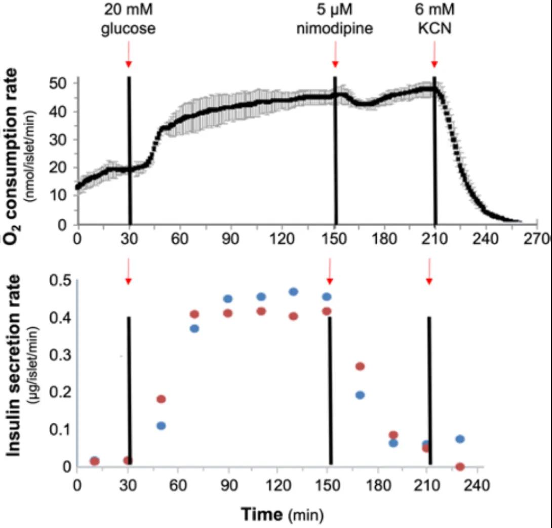
Simultaneous parallel measurements of mitochondrial respiration and insulin secretion in mouse islets. Representative measurement of oxygen consumption (top) and insulin release (bottom) from islets studied on the BaroFuse multichannel respirometry/perifusion system following infusion of glucose, nimodipine, and potassium cyanide. Top graph is the mean oxygen consumption (±SD) of two independent channels (75 islets each from islets isolated from the same mouse). Bottom graph is insulin secretion collected from the same islets from each independent well (blue, red).
Transfection
Islets can be transfected in vitro, or in vivo after I.P. delivery
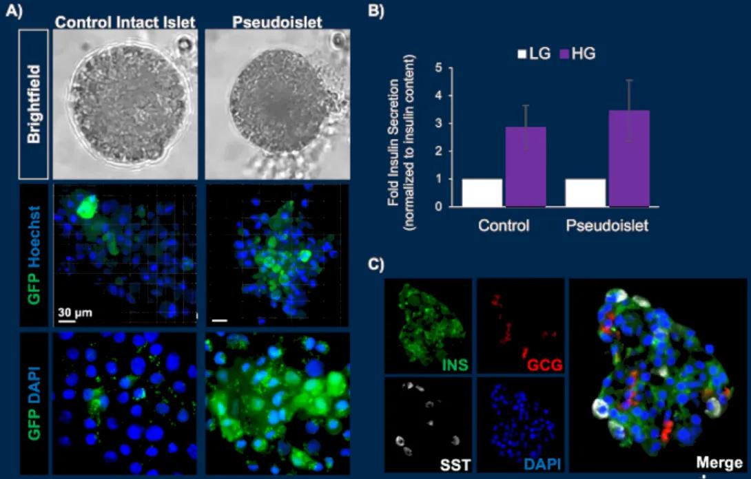
Islet dissociation and pseudo-islets
- Islet dissociation
- MACS separation of the β-cells
- Transfection of single cells
- Pseudoislet generation
- Islet isolation
- Secretion assays
- Morphometry
- Experimental design
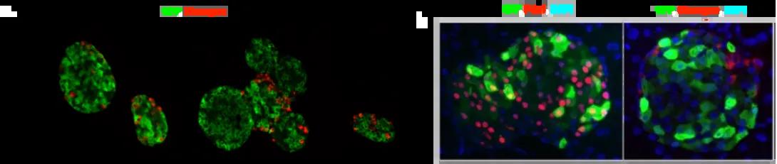
- sfGFP-CPep imaging
- Embryonic rudiments cultures
- Grafts and Imaging in the anterior chamber of the eye
Key Personnel

Scott A Soleimanpour, MD
Professor of Internal Medicine
Professor of Molecular and Integrative Physiology and Associate Director
M-Diabetes for Type I Diabetes Basic Research
Metabolism
Endocrinology and Diabetes
Medical School

Corentin Cras-Meneur
Internal Medicine
Medical School