
Microscopy & Image Analysis Core (MIAC)
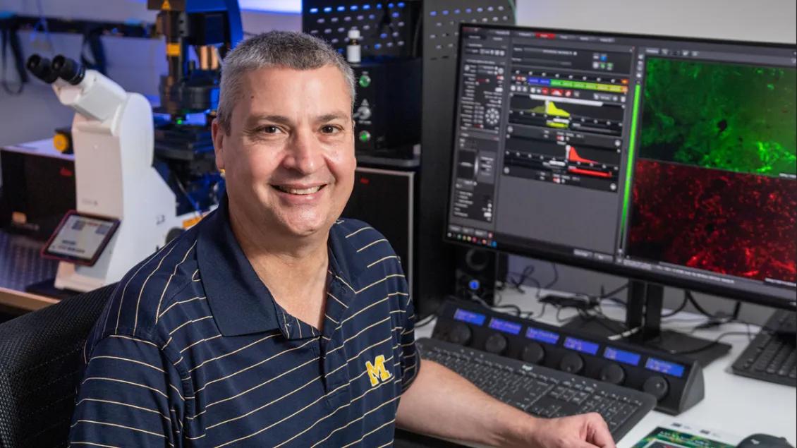
Microscopy Expertise at Your Fingertips
Located in Brehm research tower, the Microscopy and Image Analysis Core (MIAC) is a shared resource providing state-of-the-art microscopy instrumentation and image analysis software to enhance diabetes-related research at the University of Michigan that would otherwise be beyond the scope and resources of most investigators. The MIAC provides expertise, advice, training, and access to equipment for a variety of imaging modalities and image analysis techniques. The primary goal of the MIAC is to support MDRC scientists in their research focused on diabetes and its complications or related endocrine and metabolic disorders.
The MIAC is now part of the BRCF Microscopy Core. Please refer to the Microscopy Core Website for accurate, up-to-date information on our instrumentation, services, policies, and rates as this site will no longer be updated. To schedule the microscopes and image analysis workstations located at Brehm, please use the calendars on Microscopy Core MiCores site.
How We Serve Your Research Needs
Dr Lentz and other members of the Microscopy Core team are available to meet with investigators at Brehm to discuss all aspects of their imaging projects including:
- Theory and practice
- Sample preparation
- Image acquisition
- Image analysis
Microscope and Image analysis training is provided, allowing investigators to use core equipment independently.
To schedule a consultation or training, please use the web-based form.
Nikon N-SIM Superresolution + A1R Confocal
Laser Scanning Confocal, Super Resolution, Widefield
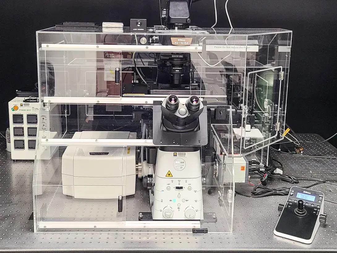
- An inverted microscope equipped with a point-scanning confocal and a 2D / 3D structured illumination (SIM) system.
- Equipped with a resonant scanner for fast confocal image acquisition.
- Time-lapse widefield live-cell imaging is also supported.
- 6 available laser lines for confocal (4 for SIM): 405nm, (445nm), 488nm, (514nm), 561nm, and 647nm.
Leica Stellaris 8 FALCON Confocal
Laser Scanning Confocal
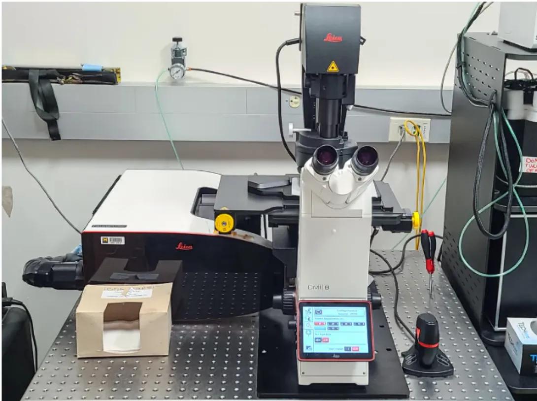
- A high-sensitivity, inverted, point-scanning confocal system, suitable for fixed and live samples.
- Equipped with a resonant scanner for fast image acquisition.
- Features FALCON for fluorescence lifetime microscopy (FLIM, includes Phasors and FCS).
- Lasers: 405nm diode laser; white light laser tunable from 440nm-790 nm.
In publications including data collected with this system, please acknowledge the NIH S10 Shared Instrumentation Grant NIH S10 OD28612-01-A1
Leica SP5 Confocal
The Leica SP5 is retiring soon. It can continue to be booked in MiCores, but no new users will be trained.
We provide several high-end image analysis workstations, which can be booked in MiCores.
We support a variety of image analysis software packages including:
- Commercial packages under support: Imaris, NIS Elements, Leica LAS and Lightning
- Commercial packages available for use but not currently under support: AutoQuant, MetaMorph, Aivia
- Open Source: Image J, Cell Profiler, QuPath, and others
Many additional resources are available through the BCRF Microscopy Core at our BSRB, MSII and North Campus Research Complex locations.
- Multiphoton Imaging
- Pixel Reassignment Superresolution
- Spinning Disk Confocal
- Lightsheet Imaging
- Tissue Clearing
- Standard sample preparation for TEM (Chemical fixation, Epon embedding and ultrathin sectioning)
- Standard sample preparation for SEM (Critical point drying and sputter coating)
- Sample Prep for Advanced Methods including FIB-SEM, Array Tomography, Immuno-EM and CLEM
- High Pressure Freezing/Freeze Substitution
- TEM and SEM imaging (training provided; staff assisted imaging also available)
For more information on the Instrumentation and Service Available at the BRCF Microscopy Core, please refer to our website.
Key Personnel
Core Director

David Antonetti
Professor of Ophthalmology and Visual Sciences
Professor of Molecular and Integrative Physiology and Associate Chair in Laboratory Research
Medical School
Lab Director
Stephen Lentz, PhD
Michigan Diabetes Research and Training Center
Diabetes I Neuropathy
Contact Us
Microscopy and Image Analysis Core Laboratory
Brehm Tower, Room 6245
1000 Wall Street
Ann Arbor, MI 48105-5714
