MIAC Equipment
Equipment for Your Research
Location
The Imaging Laboratory is in Room 6245 on the 6th floor of the Brehm Center for Diabetes Research (see maps below). The Core consists of a suite of interior rooms (6245, 6245A and 6245B) and each microscope is housed in an individual room to maximize imaging of fluorescent signals. The main area of the core consists of a laboratory with 2 benches for specimen preparation, a fume hood and space for two computer workstations and printers. In addition, the Core has access to shared space including autoclave and dish washing facilities. Tissue culture incubators are present for short-term storage of cells under a controlled environment.
The Core’s aims are to provide service, access to specialized state-of-the-art instrumentation, education and training in morphological techniques.
Confocal Microscopes
Leica Stellaris 8 Reservation Calendar + LOGIN
Quick view Leica Stellaris 8 Calendar no Login
- Leica DMi 8 Inverted Microscope
- 8 kHz Tandem Resonant Scanner
- White Light Laser -440 nm -790 nm and 405 nm Laser Diode
- AOBS (Acousto Optical Beam Splitter). Tunable crystal that allows for precise laser line excitation for up to 8 lines simultaneously
- Spectral Detectors for variable emission wavelength selection and lambda (wavelength) scanning for spectral unmixing
- 5 Power HyD Detectors (2 x HyD S, 2 x HyD X, HyD R-out to 850 nm)
- FALCON fluorescence lifetime microscopy (FLIM, includes Phasors and FCS)
- Scanning Stage SuperZ for precise control over Z scans, tiling of large areas and multi-point mark and find
- Closed Loop Focus with Adaptive Focus Control
- ToKai Hit Stage Top Incubator
- Leica LIGHTNING Super Resolution (Deconvolution)
- Software Wizards for programming specialized techniques like FRET, FRAP and photoactivation
- Located in Room 6245A Brehm Center
Publications that include data collected with the Leica STELLARIS 8 system must acknowledge support of the S10 shared instrument as follows:
“This work used a Leica STELLARIS 8 FALCON Confocal Microscopy System, funded by a National Institutes of Health SIG grant NIH S10OD28612-01-A1 and available through the Microscopy and Image Analysis Core of the Michigan Diabetes Research Center, funded by National Institutes of Health Grant P30 DK-20572 from the National Institute of Diabetes and Digestive and Kidney Diseases.”
...or...
“This work used a Leica STELLARIS 8 FALCON Confocal Microscopy System that was purchased with funds from a National Institutes of Health SIG grant NIH S10OD28612-01-A1.”
...or...
“The authors would like to acknowledge the NIH S10 Shared Instrumentation Grant NIH S10OD28612-01-A1 for supporting this work.”
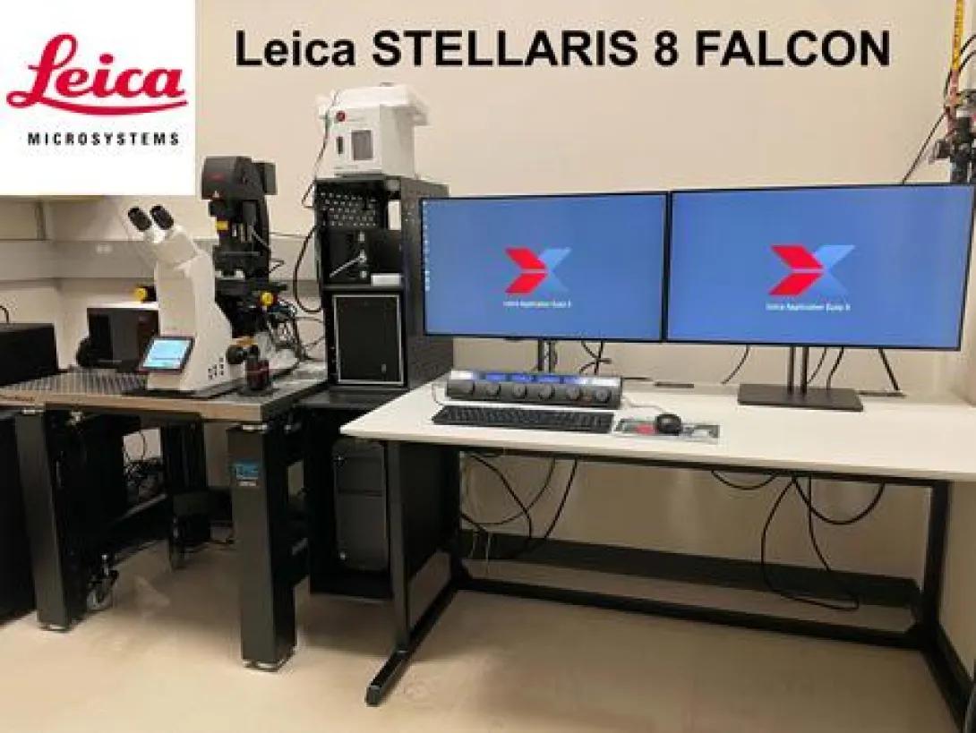
Strengths of the Leica Stellaris 8 FALCON Confocal Microscope
- 8 kHz Tandem Resonant Scanner for rapid scanning for live cell imaging or stitching large areas of tissue.
- LIGHTNING deconvolution acquisition and post processing software.
- FALCON fluorescence lifetime microscopy (FLIM, includes Phasorplotsand FCS)
- Tokai Hit environmental chamber to regulate temperature and CO2
- Software Wizards for programming specialized techniques like FRET, FRAP and photoactivation.

Nikon A1 Reservation Calendar + LOGIN
Quick view Nikon A1 Calendar no Login
- Nikon TiE Inverted Microscope
- Designed for Live Cell Imaging
- Prior motorized stage for tiling large areas and multi-point mark and find
- Nikon’s Perfect Focus System (PFS) to maintain focus on sample by compensating for thermal drifts
- Tokai Hit environmental chamber to regulate temperature and CO2
- Filter based system to direct wavelengths to the PhotoMultipler Tube detectors for confocal microscopy
- Cy-5 filter set for visualizing far red signals in the oculars/eyepieces before imaging with the laser
- Located in Room 6245B Brehm Center
Wide field Components
- Highly sensitive (95% quantum efficient) Photometrics Prime 95B cMOS monochrome camera for widefield microscopy
- Sutter filters wheels for standard blue, green and red fluorescent signals and CFP and YFP for FRET (Förster resonance energy transfer)
Strengths of the Nikon A1 Confocal Microscope
- Designed for Live Cell Imaging
- Prior motorized stage for tiling large areas and multi-point mark and find.
- Nikon’s Perfect Focus System (PFS) to maintain focus on sample by compensating for thermal drifts.
- Tokai Hit environmental chamber to regulate temperature and CO2.
- Filter based system to direct wavelengths to the PMT detectors.
- Cy-5 filter set for visualizing far red signals in the oculars/eyepieces before imaging with the laser.
Leica SP5 Reservation Calendar + LOGIN
Quick view Leica SP5 Calendar no Login
- Leica DMI 6000 Inverted Microscope
- AOBS (Acousto Optical Beam Splitter). Tunable crystal that allows for precise laser line excitation for up to 8 lines simultaneously
- Spectral Detectors for variable emission wavelength selection and lambda (wavelength) scanning for spectral unmixing
- Scanning Stage SuperZ for precise control over Z scans, tiling of large areas and multi-point mark and find
- Software Wizards for programming specialized techniques like FRET, FRAP and photoactivation
- Located in Room 7054 Brehm Center
Strengths of the Leica SP5 Confocal TCS Microscope
- AOBS (Acousto Optical Beam Splitter). Tunable crystal that allows for precise laser line excitation for up to 8 lines simultaneously.
- Spectral Detectors.
- Allows for variable emission wavelength selection.
- Lambda (wavelength) scanning for spectral unmixing.
- Scanning Stage SuperZ. Allows for precise control over Z scans, tiling of large areas and multi-point mark and find.
- Software Wizards for programming specialized techniques like FRET, FRAP and photoactivation.
Epi-Fluorescence Microscope
Nikon A1 Reservation Calendar + LOGIN
Quick view Nikon A1 Calendar no Login
- Highly sensitive (95% quantum efficient) Photometrics Prime 95B cMOS monochrome camera for widefield microscopy
- Sutter filters wheels for standard blue, green and red fluorescent signals and CFP and YFP for FRET (Förster resonance energy transfer)
- Located in Room 6245B Brehm Center
High Resolution Image Processing Workstations and Supporting Software
The Core has four high-end imaging workstations based on Windows 64-bit operating systems.
- Leica Application Suite X: Lightning Deconvolution, 3D Visualization, Fluorescence Lifetime Imaging
- Microscopy (FLIM) & phasor plots, and LIGHTNING deconvolution
- Nikon Elements
- MetaMorph version 7.10 Offline Analysis
- Imaris version 9.5: 3D and 4D Imaging
- Aivia version 12.1 - Artificial Intelligence - machine and deep learning
- AutoQuant X version X3 : 2D/3D deconvolution
- MATLAB version R2022a
- GraphPad Prism version 9.3 (Graphing & Statistical Analysis)
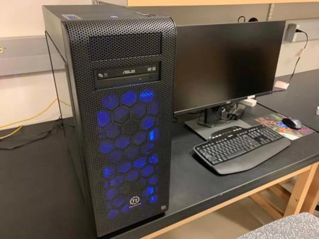
Windows PC #1 Reservation Calendar + LOGIN
Quick view Windows PC #1 Calendar no Login
Specs
- AMD 16-Core Thread ripper 3.4 GHz processor, 128 GB memory, NVIDIA Quadro P4000 8 GB graphics card, 500 and 950 GB solid state hard drives, 6 TB data drive, dual 27” Dell UP2716D 2560x1440 monitors
- Located in Room 6245 Brehm Center
Image Analysis Software
- 3D Imaris, AutoQuant deconvuloution, 3D Volocity, MATLAB, ImageJ, Offline Nikon Elements, Offline Leica Application Suite (LAS X) that includes 3D Visualization, Fluorescence Lifetime Imaging Microscopy (FLIM) & phasor plots, and LIGHTNING deconvolution, ImageJ version 1.46h: Open Source Analysis Software, Fiji version ImageJA 1.45b: Open Source Analysis Software
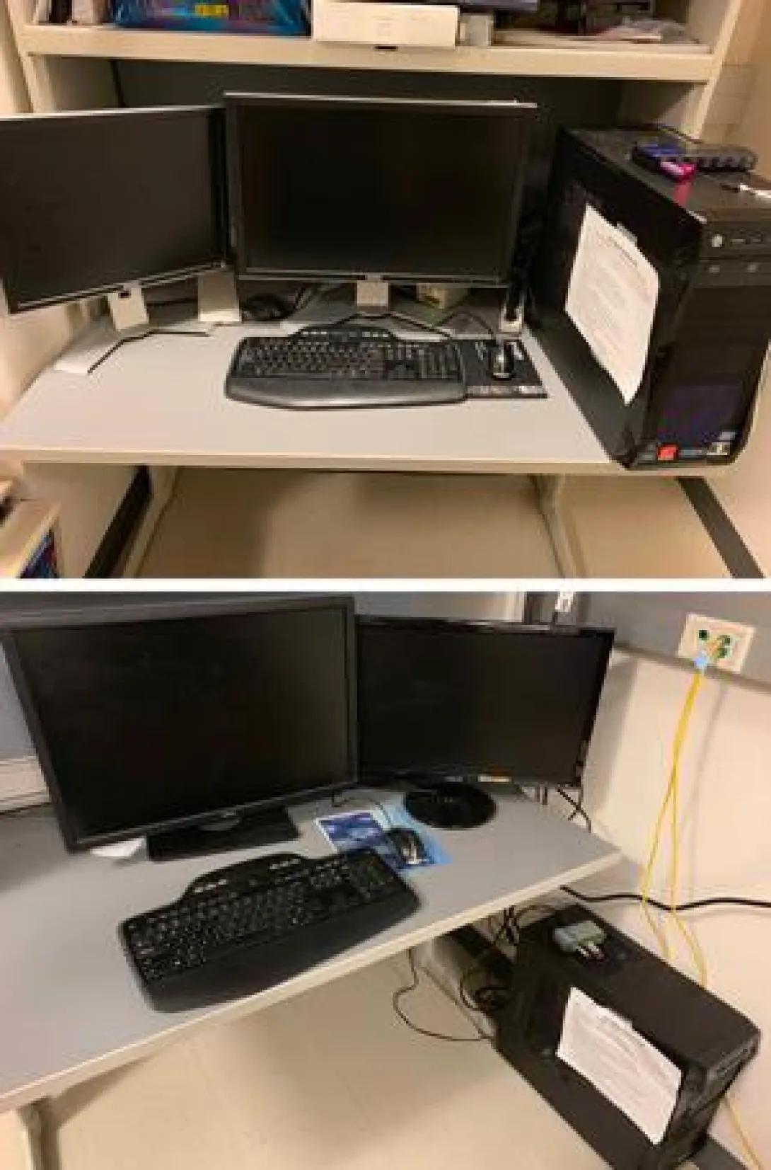
Windows PC #2 Reservation Calendar + LOGIN
Quick view Windows PC #2 Calendar no Login
Specs
- Intel i7 3.4 GHz 4-core processor, 16 GBs of RAM, Radeon HD 4650 1 GB video card and 128 GB solid state hard drive running Windows 7 32-bit or 64-bit OS, 2 TB secondary hard drive for data storage, 24" Dell UltraSharp U2408WFP 1920x1200 and 19" Dell 1907FP 1280x1024 monitors
- Located in Room 6245 Brehm Center
Image Analysis Software
- MetaMorph version 7.8.6, ImageJ version 1.51k, Fiji version ImageJA 1.5k, Microsoft Office 2016: Word, Excel, PowerPoint, Adobe Creative Suites 6 Web Design Premium: Acrobat X, PhotoShop CS5, Illustrator CS5, Dreamweaver CS5, VirtualDub 64: Video Editing Software
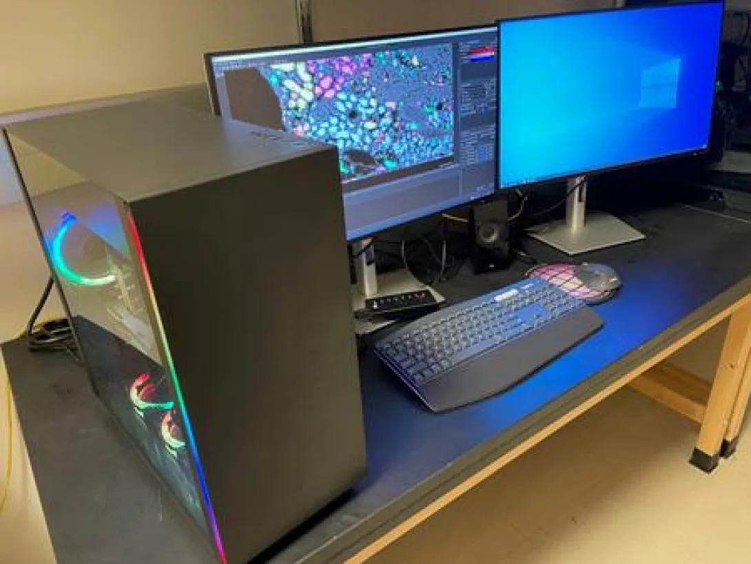
Windows PC #4 Reservation Calendar + LOGIN
Quick view Windows PC #4 Calendar no Login
Specs
- Intel Xeon 6-Core 3.8 GHz processor, 128 GB memory, NVIDIA GeForce RTX 11 GB GDDR6 graphics card, 2 TB solid state hard drives, 6 TB 7200 rpm data hard drive, two 27” Dell U2723QE 3840x2160 monitors
- Located in Room 6245 Brehm Center
Image Analysis Software
- AIVIA Artificial Intelligence-guided Software, MATLAB, Office 365
Additional High Resolution Image Processing Workstations
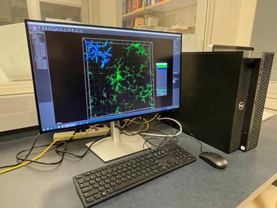
VRC Dell Precision 5820 Offline Image Analysis Reservation Calendar + LOGIN
Quick view VRC Dell Precision 5820 Offline Image Analysis no Login
Specs
- Intel Xeon 10-Core W-2255 3.7 GHz processor, 128 GB memory, Nvidia RTX A4000 16 GB 4 DP graphics card, 500 GB and 2 TB GB M.2 PCIe NVMe Class 40 solid state hard drives, 27” Dell UP2723QE 3840 x 2160 monitor
- Located in Room 7054 Brehm Center
Image Analysis Software
- 3D Imaris, MATLAB, Free Leica Application Suite (LAS X)
Specialized Equipment
The FV500 is controlled by a 2.4 Ghz personal computer under Windows XP and is capable of imaging 5 separate channels simultaneously (4 fluorescence + 1 transmitted light photomultiplier detectors) offering highly efficient, maximum emission sensitivity and the ability to record scanned images in 12 bits or 4096 gray levels, thus allowing quantitative linear measurement of fluorescence within regions of low contrast as well as very high contrast. Users are able to image a wide variety of fluorophores with laser excitation that includes Blue Violet (405nm) Multi-Line Argon Blue (458,488,515nm), Helium Neon Green (543nm) and Helium Neon Red (633nm) for standard Blue, Green, Red and Far-Red fluorochromes. The FV500’s acoustical optical tuning filter (AOTF) and adjustable scan speeds provides for minimal specimen fading, sequential scanning for reduced fluorescence cross talk, multiple regions of excitation, high resolution imaging (up to 2048 x 2048 pixels) of fixed or static samples, and rapid recording of kinetic events.
Optical sections in the z plane can be collected using a stop motor attached to the fine focus control of the microscope and driven by Fluoview software. The system is also equipped with Differential Interference Contrast (DIC) objectives and condensers and has the ability to capture transmitted light images with a highly sensitive photomultiplier (PMT) transmission detector.
Images are saved to local secondary 250 GB hard drive and then transferred to a 2 TB mirrored RAID-1 network-attached storage (NAS) device that is accessible from core workstations or individual laboratory computers through the UM medical campus 4 Gbps network. Integral Fluoview software allows for analysis of saved images in 2 dimensions (e.g. brightness vs time); confocal images obtained in a z-series can be volume rendered and analyzed in 3 dimensions. Fluoview software saves images in a non-proprietary format to facilitate the use of the raw data files in 3rd party image analysis or visualization software such as ImageJ, MetaMorph, AutoQuant, Volocity and Imaris.
Strengths of the Olympus FluoView 500 Laser Scanning Confocal Microscope
- Great work-horse system for routine 1, 2 or 3 color (blue, green, red or green, red, far red) imaging of fluorescent signals in fixed cells or tissue.
- Filter based system to direct wavelengths to the PMT detectors. In some instances signals are stronger compared to spectral detectors.
- Cy-5 filter set for visualizing far red signals in the oculars/eyepieces before imaging with the laser.
Confocal images taken by the Olympus FV500
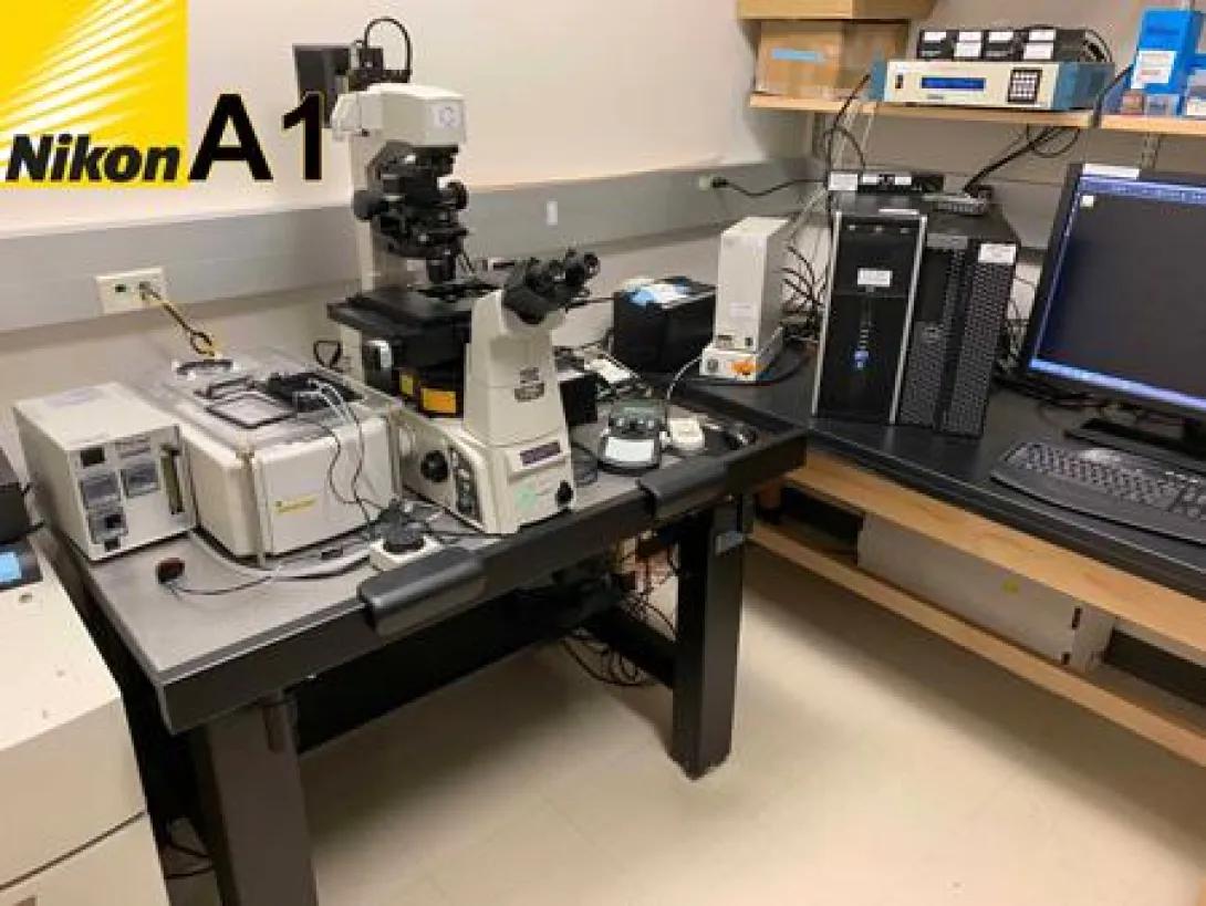
Nikon A1 Confocal Microscope
The Nikon A1 confocal combined with an inverted Ti-E microscope will accommodate both fixed and live samples. It is capable of imaging 5 separate channels (4 fluorescence + 1 transmitted light photomultiplier detectors) to capture signals from blue, green, red and far-red fluorochromes. The Nikon A1 is a filter based system to direct wavelengths to the PMT detectors and has the following laser lines to excite fluorescent signals, 405 nm laser diode, 457, 488, 514 nm argon laser, 543 nm HeNe, and 640 nm red diode. The system is controlled through Nikon’s Elements Confocal Microscopy software and is built on a HP computer with an i7 Intel 2.8 GHz CPU, 16 GB RAM running Windows 7 64-bit OS. All software and data are located on a single 1 TB hard drive and data can be transferred to the core’s 1.5 TB mirrored RAID-1 NAS.
The modular platform of the A1 system offers the greatest degree of flexibility, allowing for future upgrades to enhance the system. The Ti-E microscope was upgraded to support live cell imaging. These upgrades consisted of Nikon’s Perfect Focus System (PFS) to maintain focus on samples by compensating for thermal drifts in the microscope, a Prior motorized stage for tiling large areas and multi-point imaging and a 60x (1.2 NA) water immersion objective. A specialized stage adaptor was manufactured by the Vision Core’s machine shop to accommodate a Tokai Hit environmental chamber to regulate temperature and CO2 to support live cell imaging.
Strengths of the Nikon A1 Confocal Microscope
- Designed for Live Cell Imaging
- Prior motorized stage for tiling large areas and multi-point mark and find.
- Nikon’s Perfect Focus System (PFS) to maintain focus on sample by compensating for thermal drifts.
- Tokai Hit environmental chamber to regulate temperature and CO2.
- Filter based system to direct wavelengths to the PMT detectors.
- Cy-5 filter set for visualizing far red signals in the oculars/eyepieces before imaging with the laser.
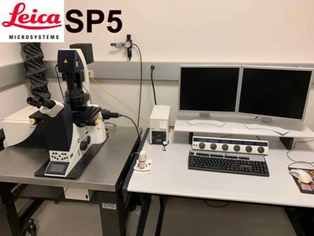
Leica SP5 Confocal TCS Microscope
The Leica TCS SP5 confocal system is shared instrument between the Diabetes and the Kellogg Eye Centers. The Leica SP5 system is housed in the Leonard G. Miller Microscopy Suite Room 7054 of the Brehm Tower. One of the strengths of the system is its AOBS Acousto Optical Beam Splitter (AOBS), a tunable crystal that allows for precise laser line excitation for up to 8 lines simultaneously, supporting excitation with the following laser lines, Laser Lines: 405 nm laser diode, 458, 476, 488, 496, 514 nm argon laser, 543 nm HeNe, 594 nm HeNe and 633 nm HeNe. The system has four spectral PMT detectors for variable emission wavelength selection and lambda (wavelength) scanning for spectral unmixing.
The DMI6000 inverted microscope has a scanning stage SuperZ for precise control over Z scans, tiling of large areas and multi-point mark and find. The Leica Application Suite Advanced Fluorescence software (version 2.5.1.6757) is easy to use and has integrated wizards for programming specialized techniques like FRET, FRAP and photoactivation. The system is controlled by a HP computer with a Xeon 3 GHz CPU and 2 GB RAM. The Windows XP professional OS and software are run on a 150 GB hard drive and data are stored on a secondary 1 TB hard drive and then transferred to the core’s 1.5 TB mirrored RAID-1 NAS. A Tokai Hit environmental chamber was purchased to regulate temperature and CO2 to support live cell imaging.
Strengths of the Leica SP5 Confocal TCS Microscope
- AOBS (Acousto Optical Beam Splitter). Tunable crystal that allows for precise laser line excitation for up to 8 lines simultaneously.
- Spectral Detectors.
- Allows for variable emission wavelength selection.
- Lambda (wavelength) scanning for spectral unmixing.
- Scanning Stage SuperZ. Allows for precise control over Z scans, tiling of large areas and multi-point mark and find.
- Tokai Hit environmental chamber to regulate temperature and CO2
- Software Wizards for programming specialized techniques like FRET, FRAP and photoactivation.
Multiwavelength Widefield Imaging System
The Imaging Laboratory has a multiwavelength widefield imaging system based on an inverted Nikon Diaphot 200 fluorescent microscope. Standard blue, green and red fluorescent signals are visualized with Chroma filter cubes. Users have the option to use either Compix SimplePCI (version 6.5.2, Hamamatsu), MetaMorph (version 7.7, Molecular Devices) or MetaFluor (version 7.7, Molecular Devices) software to capture digital images with a Hamamatsu ORCA extended range digital CCD camera (C4742-95-12ER). The system is controlled by a Dell 2.4 GHz Pentium IV personal computer with 768 MB of RAM, Matrox Meteor II digital PCI frame grabber, and an 80 GB hard drive running Windows XP operating system. Images and data can be exported in convenient formats including tiffs and Microsoft Excel spread sheets and archived on an internal CD-RW drive.
This microscope is used as our state-of-the-art fluorescence resonance energy transfer (FRET) system. This type of microscopy enables members of the MDRC to study a wide variety of biological events that influence the interaction between molecules. FRET technology involves the non-radiative transfer of energy from a fluorophore in an excited state to a nearby acceptor fluorophore. FRET has grown in popularity due to the emergence of GFP mutants with blue or yellow-shifted spectral properties. We have developed our system to take advantage of the fluorescent characteristics of enhanced cyan fluorescent protein (CFP) and yellow fluorescent protein (YFP). FRET microscopy relies on the ability to capture weak and transient fluorescent signals efficiently and rapidly from the interactions of labeled molecules in live samples. This necessitates the use of a digital camera that can perform under these stringent conditions. Our FRET system is equipped with a Hamamatsu ORCA extended range digital CCD camera (C4742-95-12ER) that can rapidly capture images at rates ranging from 8.3 - 45 frames per second with very high quantum efficiency resulting in shorter exposures of sensitive samples to fluorescent light. Thus, the ORCA digital camera allows us to get the maximum performance and utility from FRET microscopy. The acquisition and analysis of FRET data is semi-automated with the use of specialized Metamorph macros/journals developed by Dr. Joel Swanson, our FRET consultant. The customized macros also allow users the ability to perform stoichiometric FRET analysis.
This microscope is equipped with additional specialized excitation and emission filters to allow users to capture fluorescence based images of transfected green fluorescent protein chimeric molecules. The system was updated to incorporate specialized fura-2 filters into the automated Sutter filter wheels to give users the ability to measure changes in calcium in live cells. MetaFluor Ratio Fluorescence Imaging Software (version 7.7, Molecular Devices) was added to the Core’s computer to simplify acquisition and analysis. This was coordinated with the purchase of an ALA Scientific Instruments profusion system that fits our existing Warner Instruments chambers and can be used with both widefield and confocal microscopes.
Strengths of the Nikon Diaphot200 Epi-Fluorescent Microscope
- Sensitive and fast gray scale Hamamatsu ORCA extended range digital CCD camera (C4742-95-12ER).
- Ratiometric FRET Imaging and Analysis for CFP and YFP FRET Pairs.
- MetaFluor Ratio Fluorescence Imaging Software combined with FURA-2 filters.
The Core has a versatile live cell perfusion system from Warner Instruments Company (Series 20 PH-2 heated platform) with the option of having either a closed (RC-21BR) or open (RC-21BDW) perfusion chamber. Temperature is controlled automatically with a dual channel controller (TC-344B) that directly connects to the platform to monitor and regulate bath and solution temperature. Another available option is for users to grow cells in 35 mm dishes and connect to the temperature controller via a quick exchange platform (QE-1). Our component based system can be configured to work with either the Core’s confocal or wide-field fluorescent microscopes. They are interested in using a chamber system that will accommodate 35 mm dishes as well as specialized dishes.
The Core has three high-end imaging workstations based on Windows 32-bit and 64-bit operating systems. These computers support the use of image analysis software packages such as for deconvolution and 3D volumetric analysis. The Core has AutoQuant (version X2.2.2, Media Cybernetics) software that specializes in image restoration and includes a deconvolution module for producing high resolution images by using cutting-edge algorithms to perform image restoration by removing out-of-focus haze, blur, noise and other problems from both 3D and 2D images. AutoQuant also includes alignment software that will correct for any drifts or shifts in images that are acquired in time or z-series data sets. Core users also have access to Volocity (version 5.3 64-bit, Perkin Elmer) which is state-of-the-art multidimensional software to process and analyze z-series of high resolution images. The Iterative Restoration module eliminates noise and blur from the original confocal images for improvement in X, Y and Z resolution. The Visualization module provides high quality rendering in order to examine and rotate the 3D reconstructed images in real time with the detail and clarity necessary for working with volumetric data. Volocity Classification module is vital for identifying, classifying and measuring the morphological changes. The Imaging Laboratory also has Imaris software, Bitplane’s cutting-edge 3D and 4D imaging software. Imaris (version 8.0.2) allows visualization of original data objects in a real time interactive manner so investigators can quickly make visual assessments of their experiments in 3D and 4D to discover relationships that are otherwise hidden. Its rendering quality, speed, precision and interactivity are unrivalled. With a large variety of segmentation options, Imaris provides investigators with the most effective tools to segment even the toughest datasets to identify, separate, and visualize individual objects and then retrieve a comprehensive array of measurements from the objects. The software includes the ability to colocalize signals and tracking particles and link to MATLAB (v2017a, Mathworks) for specialized or customable analyses. The core continues to provide access to offline version of MetaMorph (version 7.8.6, Molecular Devices). This software provides sophisticate morphometric analysis tools and features some very useful semi-automated processes such as cell counting. These workstations also have additional analysis open source packages installed such as ImageJ (U. S. National Institutes of Health, Bethesda, Maryland), Fuji and Virtual Dub.
Specs
Intel i7-965 quad core CPU at 3.3 GHz, 12 GB RAM, an EVGA GTX 285 1024MB video card and two hard drives (a 300 GB drive at 10,000 RPM for the 64-bit OS and a 500 GB 7,200 RPM drive for the 32-bit OS), Dell UltraSharp U2408 24” Monitor 1920x1200
Software
- Leica Application Suite Offline Software (32-bit OS)
- Olympus Fluoview Offline Software (32-bit OS)
- MetaMorph version 7.6.3 Offline Analysis Software (32- or 64-bit OS)
- Imaris version 7.3: Cutting-Edge 3D and 4D Imaging Software (64-bit OS)
- Volocity version 5.3: 3D Visualization and Quantification Software (64-bit OS)
- AutoQuant X version X2.2.2: 3D deconvolution Software (64-bit OS)
- GraphPad Prism version 5.01: Graphing and Statistical Analysis Software (64-bit OS)
- Microsoft Office 2010 Pro+: Word, Excel, PowerPoint (32- or 64-bit OS)
- Adobe Creative Suites 5 Web Design Premium: Acrobat X, PhotoShop CS5, Illustrator CS5, Dreamweaver CS5 (32- or 64-bit OS)
- VirtualDub 64: Video Editing Software (64-bit OS)
- ImageJ version 1.46h: Open Source Analysis Software (32- and 64-bit OS)
- Fiji version ImageJA 1.45b: Open Source Analysis Software (32- and 64-bit OS)
Specs
Intel i7 3.4 GHz 4-core processor, 16 GBs of RAM, a Radeon HD 4650 1 GB video card and a 128 GB solid state hard drive running Windows 7 64-bit OS. The computers have secondary hard drives for data storage (2.0 TB), Dell UltraSharp U2410 24” Monitor 1920x1200
Software
- MetaMorph version 7.8.6 Offline Analysis Software
- Imaris version 8.0.2: Cutting-Edge 3D and 4D Imaging Software
- Volocity version 5.3: 3D Visualization and Quantification Software
- AutoQuant X version X2.2.2: 3D deconvolution Software
- MATLAB R2017a
- NIS-Elements AR 4.10.01 64-bit
- Leica Application Suites - Advanced Fluorescence Lite version 3.1.0
- GraphPad Prism version 6.07: Graphing and Statistical Analysis Software
- Microsoft Office 2013: Word, Excel, PowerPoint
- Adobe Creative Suites 6 Web Design Premium: Acrobat X, PhotoShop CS5, Illustrator CS5, Dreamweaver CS5
- VirtualDub 64: Video Editing Software
- ImageJ version 1.51k: Open Source Analysis Software
- Fiji version ImageJA 1.5k: Open Source Analysis Software
Image data sets are becoming increasingly larger with the advancement of detectors, computers and storage devices associated with the core’s microscopes. The Imaging Laboratory owns a network attached storage (NAS) server to improve the storage and transfer of data collected in the core. The QNAP TS-259 Pro Turbo NAS is a powerful 2-bay server that has two 2.0 TB hard drives that provide 2.0 TBs of mirrored (RAID1) storage space. Investigators are able to safely store data acquired on the core’s equipment up to 1 month and then remotely access the server to quickly and efficiently transfer their data to their laboratory computers. This unit is expandable to accommodate additional external hard drives connected to one of its four USB 2.0 or two eSATA ports.
Specs
- CPU: Intel Atom Dual-core Processor 1.80 GHz, 1GB DDRII RAM
- Powerful 2-bay server that has two 1.5 TB hard drives that provide 1.5 TBs of mirrored (RAID1) storage space
- Ethernet w/ Jumbo Frame: Gigabit LAN x2
- FTP Server: Read = 115.7 MB/sec, Write: 114.4 MB/sec
Expansion Ports
- USB: 5 x USB 2.0 port support USB printer, pen drive, USB hub, and USB UPS
- eSATA: 2 x eSATA port
The Core also has a HP OfficeJet Pro 8610 and a Xerox ColorQube 8570DN printers that allows color prints for documentation and presentation. All printers are networked and accessible from all computer workstations.
A Nikon Labphot2A microscope is available for viewing toluidine blue stained sections. The Core is also equipped with shakers, water baths, refrigerator, pH meter, tissue culture incubators, centrifuges and other equipment.
Other equipment includes a RMC MT-7 Ultramicrotome with a CR-21 Cryosectioning Attachment, and a Reichert-Jung KNIFEMAKER II. A Nikon Labphot2A microscope is available for viewing toluidine blue stained sections. The Core is also equipped with shakers, water baths, refrigerator, pH meter, tissue culture hood and incubators, centrifuges and other equipment.
The core can advise and in some cases assist investigators who wish to use imaging instrumentation outside the core. Investigators interested in these services should make special arrangements with Dr. Stephen Lentz (e-mail: [email protected] or Telephone: 734-647-8233) for more information.
The Leica Multi-photon Confocal Microscope is maintained and operated in the Department of Ophthalmology and Visual Sciences located in the adjoining Kellogg Eye Center research building next to the Brehm Center.
- DM6000 upright
- 25X water immersion
- 405 nm laser diode
- 458, 476, 488 496, 514 nm argon laser
- 543 nm HeNe
- 675 to 1100 nm tunable Newport Ti sapphire laser
- Three spectral photomultiplier tubes (PMT)
- Three NDD (non-de-scanned detectors) (2 reflected, 1 transmitted/SHG for 2-photon imaging (NDD)
- Two APD (Avalanche Photo Detector) channels for highest sensitivity
- One CCD camera for wide field image acquisition
- Tunable Ti sapphire laser for multiphoton excitation that allows for greater depth of visualization (up to 1000 µm depending on tissue). The infrared laser causes less photoxicity for live cell/tissue imaging.
- NDD and APD Detectors
- NDD (non-descanning detectors) allows for second harmonic generation imaging (SHG), SHG-signal is generated from highly ordered structures and has been used to image collagen fibres, microtubules, the striation pattern of muscles and starch granules
- APD (Avalanche Photo Detector) channels for highest sensitivity
A JEOL JSM 1400--+Transmission Electon Microscope (TEM) is maintained and operated in Microscopy and Image-analysis Laboratory, part of the Department of Cell and Developmental Biology. The JEOL JSM 1400 is equipped with a computer controlled stage and two high resolution digital cameras. AMT software drives the digital cameras and provides on screen live viewing. This microscope is used for the collection of digital EM images and for the direct transfer of data from the microscope to the image analysis software reducing the need for photographic film, chemicals, and paper. Use of this equipment is available to University of Michigan investigators on a recharge basis.